
|
|
1.IntroductionThe unique optical properties of plasmon-resonant particles together with the high specificity of biomolecular recognition opens new possibilities for applications to extrasensitive detection of different analytes,1 biomedical diagnostics and therapy,2 targeted drug delivery,3, 4 laser killing of cancer cells,5, 6, 7 and optical imaging.8, 9, 10 However, for optical imaging at the cell and tissue levels, the most popular labels are quantum dots (QD) 11, 12, 13, 14 rather than noble metal nanoparticles. The basic advantage of semiconductor nanocrystals, in comparison with plasmon-resonant (PR) noble metal particles, is in the remarkably narrow fluorescent peaks of QDs vs light-scattering plasmon resonances. The fluorescent spectra of QDs can be tuned to a desired spectral band by varying the QD size and composition, and functionalization of QDs with recognizing biomolecules allows one to produce multicolor biospecific labels for target molecules and cells. The essential drawbacks of QD labels are the need to use UV exciting light, the quenching of fluorescence caused by the adsorption of biopolymers on the QD surface, and the biological toxicity of most QDs. Furthermore, there are difficulties in the functionalization of QDs while transferring the particles from primarily the organic synthesis phase to a water-saline environment, which needs to be done with all biospecific molecular probes. Most of these difficulties may be overcome by using PR particles, but the optical properties of these particles should be tuned in a proper manner to ensure the desired resonance quality and position. In particular, the solid gold and silver spheres are poor candidates for multicolor labeling, as their size-dependent spectral tuning covers rather narrow spectral intervals15, 16 and the spectral width of the PR scattering resonance of gold spheres is about . Among the numerous particle structures made available through the existing synthetic technologies,17 the gold nanoshells are of significant interest for applications to biosensorics,18 immunoassays,19 laser phototherapy of cancer,20, 21, 22 and optical imaging.7 The silica/gold nanoshells can easily be tuned to a desired spectral band23 from VIS to NIR. However, a typical full width at half-maximum (FWHM) of silica/gold nanoshell spectra is about , which enables one to use only three or four distinct biomarkers in the whole VIS-NIR range. Recently, Chen 24 reported on a theoretical investigation of three-layer nanoparticles with a metal core, an intermediate silica layer, and a surface metal shell. Such structures, in the authors’ opinion, may be used as multicolor labels for spectral tissue imaging. The principal result obtained by Chen 24 is that the three-layer metal-dielectric-metal (MDM) nanoparticles possess ultrasharp light-scattering resonances, which can be tuned from VIS to NIR spectral bands by varying the structural MDM parameters. By contrast with silica/gold nanoshells, the spectral widths of MDM resonances were found24 to be weakly dependent on particle polydispersity—a property that could be important for applications of MDM multicolor labels in light-scattering spectroscopic imaging, optical coherent tomography, and other fields. However, the theoretical computations of scattering spectra in Ref. 24 were carried out without taking into account the size-dependent correction of dielectric functions of thin metal shells. It is well known15 that various mechanisms contribute to the bulk dielectric function of metals on a nanometer scale,25 with the size-limiting correction26, 27 being the major factor in the case of two-layer metal nanoshells. 25, 28, 29 Here we show that the size-corrected dielectric function is a crucial factor that limits the actual widths of the scattering plasmon resonances for three-layer MDM structures. Furthermore, contrary to Chen ’s data, we did not find any significant advantages of three-layer MDM structures in comparison with the usual silica/metal nanoshells, because the size-corrected absolute FWHM of both types of particles turned out to be similar. Thus, although several attempts at the improving MDM nanoparticle synthesis have been reported,30, 31, 32 their optical advantages seem disputable at least. 2.MethodsWe consider MDM nanoparticles (external diameter ) embedded in a dielectric medium with the refractive index (in this case, water). The normalized scattering cross section, , was calculated by the well-known formulae,15 in which the usual Mie coefficients of a homogeneous sphere, and , were replaced by the corresponding coefficients of a multilayer sphere, and ( is the number of layers). To calculate the coefficients and , we used the effective recursive algorithm of Wu and Wang,33 which had previously been implemented for the case of gold/polymer conjugates.34, 35 For thin gold or silver nanoshells, the dielectric function of bulk metal should be corrected for the size-limiting effects, which restrict the mean free path of conductive electrons (for a review, see, e.g., Refs. 26, 27). A thorough comparison of the experimental and calculated colloidal gold spectra16, 36 showed that only the imaginary part of the bulk metal permittivity should be corrected: where is the effective mean free path of electrons, is the wavelength of plasma oscillations, and and are the Fermi velocity of electrons and the light velocity in vacuum, respectively. The dimensionless parameter is determined by details of the electron scattering process25, 27 and is often assumed to be close to 1. The classic effective mean free path of electrons in spherical particles varies from (isotropic scattering) to (diffusive scattering).27 The size-limiting effects in metal nanoshells, unlike those in homogeneous particles of various shape,27, 37 have not been studied with proper exactness (see, however, a series of papers by Prodan and Nordlander38, 39, 40 on the first-principle calculations). In this work, we use a formula derived by Kachan and Ponyavina29 for the effective mean free path of electrons in the case of isotropic scatteringwhere and are the outer radius and the shell thickness, respectively. Granqvist and Hunderi in their pioneering work28 used the following analytical expression for :to simulate optical properties of a glass containing silver particles with dielectric cores by using the Maxwell Garnet rule and the dipole polarizability of core/shell particles.15 For thick shells, Eqs. 2, 3 give close results , whereas, for thin shells Eq. 3 gives overestimated -values.The spectral dependence of the bulk gold refractive index was obtained from a spline by analogy with Ref. 16. Specifically, for wavelengths of 210.4– , the spline nods were taken from Irani; 41 for those of , from Otter;42 and for those of , from Johnson and Christy.43 The bulk refractive index of silver was obtained from Johnson and Christy’s43 data. 3.Results and DiscussionAll the nanostructures examined in this paper correspond to those studied by Chen 24 Figure 1 shows the scattering spectra of solid gold nanospheres, two-layer structures, and three-layer structures. The spectra were calculated by using size-corrected dielectric functions (solid lines) and bulk optical constants (dashed lines). Evidently, the size-limiting correction of the optical constants leads to dramatic changes in the FWHM values and in the absolute maxima, whereas the resonance spectral positions coincide in both cases. We emphasize that the size-limiting correction of the optical constants is an important factor that allows one to bring the measured and calculated spectra into satisfactory agreement for suspensions of solid spheres16, 44 and gold nanoshells25 as well as for single spherical particles.45 On the other hand, it has been reported that the calculations with the bulk dielectric function reproduce the experimental light scattering spectra of individual gold nanoshells46 and nanorods.47 Thus, one needs further studies of the issue because the nanorod size and shell thickness in the cited reports46, 47 were too large for noticeable size-correction effects. Fig. 1Comparison of the resonant light-scattering peaks for solid, two-layer, and three-layer nanospheres. The solid curves correspond to calculations with a size-corrected Au dielectric function, and the dashed curves were calculated with bulk optical constants. The sphere structure is designated in the legend as (core radius)/(core material); (thickness of shell)/(material of shell). All dimensions are in nanometers. Here and in what follows Au, Ag, and Sil stand for gold, silver, and silica, respectively. 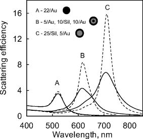 Following the argumentation in Ref. 24, we discuss further the optical properties of MDM structures only in terms of the normalized (to the resonance peak value) scattering spectra and FWHM, without consideration of the absolute resonance values. In accord with earlier experimental observations and theoretical simulations,17, 23 Fig. 2 shows the normalized scattering spectra of two-layer particles that are tuned to the VIS-NIR resonance position by varying the core/shell ratio at a constant external diameter of and at a constant dielectric/metal composition. Note that our noncorrected spectra are in full agreement with the data of Chen 24 However, after appropriate correction for the size-limiting effects, all spectra become essentially broadened and their peak-to-baseline ratio approaches the range from 3 to 5, which is close to that of the solid nanosphere resonances. In the case of three-layer MDM structures (Fig. 3 ), we obtained similar results; therefore, no additional comments are needed. Fig. 2Resonant light-scattering peaks of two-layer nanospheres with silica cores calculated without (a) and with (b) a size correction of the dielectric function. The overall radius of nanostructures with a variable relative dimension of the core versus the shell is fixed . The dielectric/metal structure is designated in the legend as (core radius)/(core material), (thickness of the shell)/(material of the shell). All dimensions are in nanometers.  Fig. 3Resonant light-scattering peaks of three-layer nanospheres calculated without (a) and with (b) a size correction of the dielectric function. The overall radius of nanostructures with a variable relative dimension of the core versus the shells is fixed . The MDM structure is designated in the legend as (core radius)/(core material), (thickness of shell 1)/(material of shell 1), (thickness of shell 2)/(material of shell 2). All dimensions are in nanometers.  Figure 4 presents a summary of Figs. 2 and 3 in terms of the FWHM values for structures designated by the letters A–J in Figs. 2 and 3. The solid and gray columns correspond to the noncorrected and size-corrected calculations, respectively. The corrected FWHM values lie within the range of , depending on the resonance peak position. Clearly, these resonance widths are close to the solid-sphere FWHM values. Fig. 4Dependence of the FWHMs of the resonance peaks of two- (a) and three-layer (b) nanoshells. The gray area corresponds to the data from calculations using according to Eq. 2. The solid area corresponds to calculations without a size-limiting correction. Denotement from A to J corresponds to the structures shown in Fig. 2a and Fig. 3b.  4.ConclusionsIn this work, we have examined the same dielectric/metal and MDM spherically symmetric nanostructures that were studied previously by Chen 24 The only difference between the two studies was the use of bulk or size-corrected dielectric functions for metal shells, whereas the computational algorithm was based on the same recurrence scheme, suggested by Wu and Wang.33 We have shown that scattering of electrons at the metal shell surface is accomplished by a decrease in the resonance peak value and by a dramatic increase in the FWHM values. By contrast with the previous conclusions,24 we did not find any significant differences in the scattering resonances of the usual silica/metal nanoshells and three-layer MDM nanostructures. We did not consider particle-size polydispersity because it is clear without any calculations that the resonance scattering spectra of a polydisperse sample is simply the sum of monodisperse spectra weighed with a particular fraction content. Evidently, polydispersity will result in the well-known, size-dependent broadening of spectra, caused by different resonance positions of different species. To summarize, we have not found any significant advantages of three-layer MDM spherical structures for applications to OCT and to light-scattering spectroscopy imaging. AcknowledgmentThis work was partly supported by grants from RFBR (No. 05-02-16776a) and Analytical Targeting Program “The Development of Scientific Potential of High School (2006–2008),” project RNP.2.1.1.4473. BK was supported by grants from CRDF (BRHE Annex BF4M06 Y2-B-06-08) and from the President of the Russian Federation (No. MK 961.2005.2). We thank D. N. Tychinin (IBPPM RAS) for his help in preparation of the manuscript. ReferencesD. A. Stuart,
A. J. Haes,
C. R. Yonzon,
E. M. Hicks, and
R. P. Van Duyne,
“Biological applications of localised surface plasmonic phenomenae,”
IEE Proc. Nanobiotechnol., 152 13
–32
(2005). Google Scholar
T. Kubik,
K. Bogunia-Kubik, and
M. Sugisaka,
“Nanotechnology on duty in medical applications,”
Curr. Pharm. Biotechnol., 6 17
–33
(2005). 1389-2010 Google Scholar
G. F. Paciotti,
L. Myer,
D. Weinreich,
D. Goia,
N. Pavel,
R. E. McLaughlin, and
L. Tamarkin,
“Colloidal gold: A novel nanoparticle vector for tumor directed drug delivery,”
Drug Delivery, 11 169
–183
(2004). Google Scholar
T. Pellegrino,
S. Kudera,
T. Liedl,
A. M. Javier,
L. Manna, and
W. J. Parak,
“On the development of colloidal nanoparticles towards multifunctional structures and their possible use for biological applications,”
Small, 1 49
–60
(2005). 1613-6810 Google Scholar
C. M. Pitsillides,
E. K. Joe,
X. Wei,
R. R. Anderson, and
C. P. Lin,
“Selective cell targeting with light-absorbing microparticles and nanoparticles,”
Biophys. J., 84 4023
–4032
(2003). 0006-3495 Google Scholar
V. Zharov,
V. Galitovsky, and
M. Viegas,
“Photothermal detection of local thermal effects during selective nanophotothermolysis,”
Appl. Phys. Lett., 83 4897
–4899
(2003). https://doi.org/10.1063/1.1632546 0003-6951 Google Scholar
L. R. Hirsch,
R. J. Stafford,
J. A. Bankson,
S. R. Sershen,
B. Rivera,
R. E. Price,
J. D. Hazle,
N. J. Halas, and
J. L. West,
“Nanoshell-mediated near-infrared thermal therapy of tumors under magnetic resonance guidance,”
Proc. Natl. Acad. Sci. U.S.A., 23 13549
–13554
(2003). 0027-8424 Google Scholar
C. Loo,
L. Hirsch,
M. Lee,
E. Chang,
J. West,
N. Halas, and
R. Drezek,
“”Gold nanoshell bioconjugates for molecular imaging in living cells,”
Opt. Lett., 30 1012
–1014
(2005). https://doi.org/10.1364/OL.30.001012 0146-9592 Google Scholar
J. Chen,
F. Saeki,
B. J. Wiley,
H. Cang,
M. J. Cobb,
Z.-Y. Li,
L. Au,
H. Zhang,
M. B. Kimmey,
X. Li, and
Y. Xia,
“Gold nanocages: Bioconjugation and their potential use as optical imaging contrast agents,”
Nano Lett., 5 473
–477
(2005). https://doi.org/10.1021/nl047950t 1530-6984 Google Scholar
I. H. El-Sayed,
X. Huang, and
M. A. El-Sayed,
“Surface plasmon resonance scattering and absorption of anti-egfr antibody conjugated gold nanoparticles in cancer diagnostics: Applications in oral cancer,”
Nano Lett., 5 829
–834
(2005). 1530-6984 Google Scholar
J. K. Jaiswal,
H. Mattoussi,
J. M. Mauro, and
S. M. Simon,
“Long-term multiple color imaging of live cells using quantum dot bioconjugates,”
Nat. Biotechnol., 21 32000
–32003
(2003). 1087-0156 Google Scholar
M. Y. Han,
X. H. Gao,
J. Z. Su, and
S. M. Nie,
“Quantum-dot-tagged microbeads for multiplexed optical coding of biomolecules,”
Nat. Biotechnol., 19 631
–635
(2001). https://doi.org/10.1038/90228 1087-0156 Google Scholar
W. C. W. Chan,
D. J. Maxwell,
X. Gao,
R. E. Bailey,
M. Han, and
S. Nie,
“Luminescent quantum dots for multiplexed biological detection and imaging,”
Curr. Opin. Biotechnol., 13 40
–46
(2002). https://doi.org/10.1016/S0958-1669(02)00282-3 0958-1669 Google Scholar
B. Ballou,
B. C. Lagerholm,
L. A. Ernst,
M. P. Bruchez, and
A. S. Waggoner,
“Noninvasive imaging of quantum dots in mice,”
Bioconjugate Chem., 15 79
–86
(2004). https://doi.org/10.1021/bc034153y 1043-1802 Google Scholar
C. F. Bohren and
D. R. Huffman, Absorption and Scattering of Light by Small Particles, Wiley, New York (1983). Google Scholar
N. G. Khlebtsov,
V. A. Bogatyrev,
L. A. Dykman, and
A. G. Melnikov,
“Spectral extinction of colloidal gold and its biospecific conjugates,”
J. Colloid Interface Sci., 180 436
–445
(1996). https://doi.org/10.1006/jcis.1996.0323 0021-9797 Google Scholar
Y. Xia and
N. J. Halas,
“Shape-controlled synthesis and surface plasmonic properties of metallic nanostructures,”
MRS Bull., 30 338
–348
(2005). 0883-7694 Google Scholar
J. L. West and
N. J. Halas,
“Engineered nanomaterials for biophotonics application: improving sensing, imaging, and therapeutics,”
Annu. Rev. Biomed. Eng., 5 285
–292
(2003). https://doi.org/10.1146/annurev.bioeng.5.011303.120723 1523-9829 Google Scholar
L. R. Hirsch,
J. B. Jackson,
A. Lee,
N. J. Halas, and
J. L. West,
“A whole blood immunoassay using gold nanoshells,”
Anal. Chem., 75 2377
–2381
(2003). https://doi.org/10.1021/ac0262210 0003-2700 Google Scholar
L. R. Hirsch,
R. J. Stafford,
J. A. Bankson,
S. R. Sershen,
B. Rivera,
R. E. Price,
J. D. Hazle,
N. J. Halas, and
L. J. West,
“Nanoshell-mediated near-infrared thermal therapy of tumors under magnetic resonance guidance,”
Proc. Natl. Acad. Sci. U.S.A., 23 13549
–13555
(2003). 0027-8424 Google Scholar
C. Loo,
L. Hirsch,
M. Lee,
E. Chang,
J. West,
N. J. Halas, and
R. Drezek,
“Gold nanoshell bioconjugates for molecular imaging in living cells,”
Opt. Lett., 30 1012
–1014
(2005). https://doi.org/10.1364/OL.30.001012 0146-9592 Google Scholar
C. Loo,
A. Lin,
L. Hirsch,
M. H. Lee,
J. Barton,
N. Halas,
J. West, and
R. Drezek,
“Nanoshell-enabled photonics-based imaging and therapy of cancer,”
Technol. Cancer Res. Treat., 3 33
–40
(2004). 1533-0346 Google Scholar
N. Halas,
“Playing plasmons: Tuning the optical resonant properties of metallic nanoshells,”
MRS Bull., 30 362
–367
(2005). 0883-7694 Google Scholar
K. Chen,
Ya. Liu,
G. Ameer, and
V. Backman,
“Optimal design of structured nanospheres for ultrasharp light-scattering resonances as molecular imaging multilabels,”
J. Biomed. Opt., 10
(2), 024005
(2005). https://doi.org/10.1117/1.1899684 1083-3668 Google Scholar
S. L. Westcott,
J. B. Jackson,
C. Radloff, and
N. J. Halas,
“Relative contributions to the plasmon line shape of metal nanoshells,”
Phys. Rev. B, 66 155431
(2002). https://doi.org/10.1103/PhysRevB.66.155431 0163-1829 Google Scholar
M. Quinten,
“Optical constants of gold and silver clusters in the spectral range between and ,”
Z. Phys. B, 101 211
–217
(1996). https://doi.org/10.1007/s002570050202 0340-224X Google Scholar
E. A. Coronado and
G. C. Schatz,
“Surface plasmon broadening for arbitrary shape nanoparticles: A geometrical probability approach,”
J. Chem. Phys., 119 3926
–3934
(2003). https://doi.org/10.1063/1.1587686 0021-9606 Google Scholar
C. G. Granqvist and
O. Hunderi,
“Optical absorption of ultrafine metal spheres with dielectric cores,”
Z. Phys. B, 30 47
–51
(1978). https://doi.org/10.1007/BF01323667 0340-224X Google Scholar
S. M. Kachan and
A. N. Ponyavina,
“Resonance absorption spectra of composites containing metalcoated nanoparticles,”
J. Mol. Struct., 267 563
–564
(2001). 0022-2860 Google Scholar
L. M. Liz-Marzán,
M. Giersig, and
P. Mulvaney,
“Synthesis of nanosized gold-silica core-shell particles,”
Langmuir, 12 4329
–4335
(1996). https://doi.org/10.1021/la9601871 0743-7463 Google Scholar
F. Caruso,
M. Spasova,
V. Salgueirino-Maceira, and
L. M. Liz-Marzán,
“Multilayer assemblies of silica-encapsulated gold nanoparticles on decomposable colloid templates,”
Adv. Mater. (Weinheim, Ger.), 13 1090
–1094
(2001). https://doi.org/10.1002/1521-4095(200107)13:14<1090::AID-ADMA1090>3.0.CO;2-H 0935-9648 Google Scholar
M. Ming,
Y. Chen, and
A. Katz,
“Synthesis and characterization of gold-silica nanoparticles incorporating a mercaptosilane core-shell interface,”
Langmuir, 18 8566
–8572
(2002). https://doi.org/10.1021/la026055r 0743-7463 Google Scholar
Z. C. Wu and
Y. P. Wang,
“Electromagnetic scattering for multilayered sphere: Recursive algorithms,”
Radio Sci., 26 1393
–1401
(1991). 0048-6604 Google Scholar
N. G. Khlebtsov,
V. A. Bogatyrev,
L. A. Dykman,
B. N. Khlebtsov, and
P. Englebienne,
“A multilayer model for gold nanoparticle bioconjugates: Application to study of gelatin and human IgG adsorption using extinction and light scattering spectra and the dynamic light scattering method,”
Colloid J., 65 622
–635
(2003). 1061-933X Google Scholar
N. G. Khlebtsov,
“Optical models for conjugates of gold and silver nanoparticles with biomacromolecules,”
J. Quant. Spectrosc. Radiat. Transf., 89 143
–152
(2004). 0022-4073 Google Scholar
L. B. Scaffardi,
N. Pellegri,
O. de Sanctis, and
J. O. Tocho,
“Sizing gold nanoparticles by optical extinction spectroscopy,”
Nanotechnology, 16 158
–163
(2005). https://doi.org/10.1088/0957-4484/16/1/030 0957-4484 Google Scholar
S. Bruzzone,
G. P. Arrighini, and
C. Guidotti,
“Some spectroscopic properties of gold nanorods according to a schematic quantum model founded on the dielectric behavior of the electron-gas confined in a box. I,”
Chem. Phys., 291 125
–140
(2003). 0301-0104 Google Scholar
E. Prodan and
P. Nordlander,
“Electronic structure and polarizability of metallic nanoshells,”
Chem. Phys. Lett., 352 140
–146
(2002). https://doi.org/10.1016/S0009-2614(01)01409-9 0009-2614 Google Scholar
E. Prodan and
P. Nordlander,
“The effect of a dielectric core and embedding medium on the polarizability of metallic nanoshells,”
Chem. Phys. Lett., 360 325
–332
(2002). https://doi.org/10.1016/S0009-2614(02)00850-3 0009-2614 Google Scholar
E. Prodan and
P. Nordlander,
“Structural tunability of the plasmon resonances in metallic nanoshells,”
Nano Lett., 3 543
–547
(2003). https://doi.org/10.1021/nl034030m 1530-6984 Google Scholar
G. B. Irani,
T. Huen, and
F. Wooten,
“Optical constants of silver and gold in the visible and vacuum ultraviolet,”
J. Opt. Soc. Am., 61 128
–129
(1971). 0030-3941 Google Scholar
M. Otter,
“Optische konstanten massiver metalle,”
Z. Phys., 161 163
–178
(1961). 0044-3328 Google Scholar
P. B. Johnson and
R. W. Christy,
“Optical constants of noble metals,”
Phys. Rev. B, 6 4370
–4379
(1972). https://doi.org/10.1103/PhysRevB.6.4370 0163-1829 Google Scholar
L. B. Scaffardi and
J. O. Tocho,
“Size dependence of refractive index of gold nanoparticles,”
Nanotechnology, 17 1309
–1315
(2006). 0957-4484 Google Scholar
S. Berciaud,
L. Cognet,
P. Tamarat, and
B. Lounis,
“Observation of intrinsic size effects in the optical response of individual gold nanoparticles,”
Nano Lett., 5 515
–518
(2005). https://doi.org/10.1021/nl050062t 1530-6984 Google Scholar
C. L. Nehl,
N. K. Grady,
G. P. Goodrich,
F. Tam,
N. J. Halas, and
J. H. Hafner,
“Scattering spectra of single gold nanoshells,”
Nano Lett., 12 2355
–2359
(2005). 1530-6984 Google Scholar
C. Sönnichsen,
T. Franzl,
T. Wilk,
G. von Plessen,
J. Feldmann,
O. Wilson, and
P. Mulvaney,
“Drastic reduction of plasmon damping in gold nanorods,”
Phys. Rev. Lett., 88 077402
(2002). https://doi.org/10.1103/PhysRevLett.88.077402 0031-9007 Google Scholar
|


