
|
|
1.IntroductionAnimal cell cultivation is a very important technology for pharmaceutical production and cell processing for regenerative medicine. To optimize and control the quality of cells during cultivation, monitoring of cell quantity and quality can be carried out nondestructively and noninvasively, especially in cell processing for transplantation and regenerative medicine. Although light microscopic observation is useful for the noninvasive monitoring of adhered animal cells, this technique cannot facilitate 3-D morphological observation, but only 2-D morphological observation. An atomic force microscope (AFM) can be used to observe the 3-D morphology of adhered animal cells. For example, the differences in 3-D morphology between Chinese hamster ovary (CHO) cells cultivated under various osmolarities were measured under AFM after fixation treatment of cells.1 Because 3-D observation of adhered animal cells using AFM requires a long time and fixing treatment of cells, AFM observation is considered invasive for cells. Recently, a novel phase-shifting laser microscope (PLM) was developed.2 A biprism, located between the magnifying lens and the observation plane, was used as a beamsplitter. (Fig. 1 ) The biprism was laterally translated to introduce phase shifts required for quantitative phase measurement with a phase-shifting technique. Namely, the phase shift caused by the difference in refractive indices between the sample and the reference expressed in Eq. 1 can be determined using PLM. where is the phase shift (-); is the thickness of the sample (nanometers); is the wavelength of the laser (nm); and and are the refractive indices of the sample and reference (-), respectively. We believe that PLM may be applicable for measuring the thickness of living adhered cells by substituting the refractive indices of cells and the medium to and in Eq. 1, respectively.In this study, noninvasive 3-D observation of animal cells under PLM without fixing treatment was investigated. 2.Materials and Methods2.1.Cells and MediaTissue plasminogen activator (tPA)-producing Chinese hamster ovary (CHO) 1-15500 cells (ATCC CRL-9606) were used. Ham’s F-12K medium (Dainippon Seiyaku Company, Limited, Japan) supplemented with 10% newborn bovine serum (NBS, Gibco, New York), streptomycin , and penicillin was employed for cell growth. Medium osmolarity was adjusted to 300 and by the addition of solution . Medium osmolarity was determined by measuring the depression of the freezing point using an osmometer (model OM-801, Vogel, Germany). CHO cells ( cells) were inoculated into of the medium in a dish (Falcon, 9.6 cm2) and incubated for 4 d in a incubator ( , 5% ). Cell suspension was prepared by trypsinization of adhered cells on a dish. Human bone marrow mesenchymal stem cells (MSCs) were isolated from bone marrow aspirate obtained by routine iliac crest aspiration from human donors (age 65 to 73) as previously reported.3 The content of cells among the cells analyzed by a flowcytometer was approximately 90% (data not shown).4 MSCs were cultured at densities of employing DMEM-LG (Gibco, New York) supplemented with 10% Fetal Calf Serum (FCS) (Gibco), penicillin, and streptomycin. Human umbilical cord vein endothelial cells (HUVEC) were purchased from Cambrex Bio Science Walkersville, Incorporated (Maryland) and cultured at densities of employing DMEM (Gibco, Japan) supplemented with 10% inactivated fetal bovine serum (Gibco), 1 aliquot (of the same concentration as that in MEM) of MEM nonessential amino acids (Gibco), 2-mM glutamine, penicillin, and streptomycin (Sigma, Missouri). 2.2.Cell Morphology Analysis under Phase-Shifting Laser MicroscopeThe adhered cells on the bottom surface of the culture dish were observed under PLM. Two neighboring fields of view with cells and without cells were selected as sample and reference fields, respectively, and phase shift was determined for all pixels in the sample field under PLM. By substituting the wavelength of the laser , the refractive indices of the cells determined by the method mentioned later ( , 1.375, and 1.375 for CHO, MSC, and HUVEC, respectively) and the medium , and the measured phase shift to Eq. 2, the height of the cells was calculated for all pixels in the sample field, and a 3-D view of the sample field was made. 2.3.Determination of Refractive Index of CellsThe culture medium was replaced with saline containing different concentrations of bovine serum albumin (380 to ), in which osmolarity was adjusted to the original medium osmolarity, and cells were observed under PLM. The refractive index of the saline, with which cell images disapperared under PLM, was considered as the refractive index of the cells. The refractive indices of the medium and saline were determined using a refract meter (DR-A1, Atago Company, Tokyo, Japan). 2.4.Cell Observation under Atomic Force MicroscopeCells were fixed with glutaraldehyde (4%), dried, and analyzed using an atomic force microscope (AFM)(NanoScope IIIa, Veeco). Namely, the height was scanned for all parts of each cell, and the closed area showing positive height was defined as the cell adhesive area.5 3.Results and Discussion3.1.Determination of Refractive Index of Adhered Chinese Hamster Ovary CellsAfter the CHO cells in the culture medium were observed under PLM, the culture medium was replaced with saline containing albumin while the culture dish was fixed on the stage of PLM. Cell image was distinct with the saline, having refractive indices of 1.33 and 1.34 (Fig. 2 ). Cell image was indistinct for the saline refractive index of 1.38 and was not observed for the saline refractive index of 1.39. Consequently, the refractive index of the observed CHO cells was determined to be 1.39. Fig. 2Determination of refractive index of adhered CHO cells. CHO cells adhered to a culture dish containing saline with known refractive indices (a) 1.33, (b) 1.34, (c) 1.38, and (d) 1.39 were observed under PLM. The color bar is analogous to that in Fig. 3.  Fig. 3Observation of CHO cells cultivated under different osmolarities under PLM. 3-D views of CHO cells cultivated under (a) 300 and (b) . 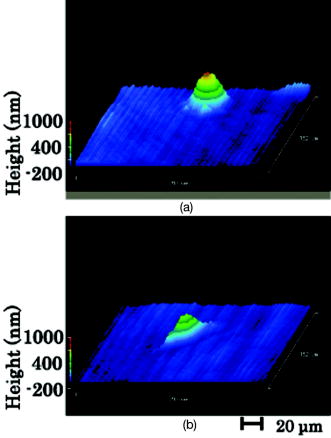 The refractive index of the cell membrane (1.5) is higher than that of the cytoplasm (1.35),6 and the thickness of the cell membrane is approximately .7 Because the adopted refractive index for CHO cells in this study (1.39) was between 1.35 and 1.5, it may be the sum of the refractive indices of the cell membrane and cytoplasm. However, the digit of significant figures must increase to increase the accuracy of the cell height measurement under PLM in the future, because the difference in refractive index between the cells and the medium was small (e.g., 0.05). 3.2.Effect of Osmolarity on Three-Dimensional Cell Morphology Observed under Phase-Shifting Laser MicroscopeCHO cells cultivated for 4 under different osmolarities of 300 and were observed under PLM [Figs. 3a and 3b ]. The 2-D morphology of the CHO cells cultivated under was elongated, while that of the CHO cells cultivated under 300 mOsm/L was globular. This difference in the 2-D cell morphology between the CHO cells cultivated under different osmolarities is in agreement with previous observations not only under AFM, but also under a conventional light-inverted microscope.1 The height of the CHO cells cultivated under [Fig. 3b] was lower than that of the CHO cells cultivated under [Fig. 3a]. The comparison of the average value for each ten cells showed that the cell maximum height under was apparently lower than that under (Fig. 4 ). This dependency of cell height on osmolarity is in agreement with a previous report observed under AFM.1 Moreover, there was no marked difference between the measured values under PLM and AFM (Fig. 4). These results strongly suggest the accuracy of cell height measurement using PLM. Fig. 4Effect of osmolarities on maximum cell height. The maximum height of each cell ( , ) observed under PLM (circle) was plotted against osmolarity together with that of each cell observed under AFM (square).  Fig. 5Influence of laser irradiation on 3-D cell morphology of CHO cells. The time lengths of laser irradiation for CHO cells before observation under PLM were (a) 0, (b) 5, and (c) , respectively. The color bar is analogous to that in Fig. 3 (color online only). 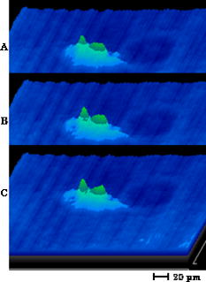 Table 2Observation of nonadherent CHO cells under PLM. Diameter and maximum height of nonadherent spherical CHO cells were measured by the observation under PLM and inverted phase contrast microscope. Mean±SD (n=10) are shown.
3.3.Comparison of Maximum Cell Heights Measured by Phase-Shifting Laser Microscope and Atomic Force Microscope for Various Cell SpeciesTo confirm the accuracy of measured cell height under PLM and the applicability of PLM to various kinds of animal cells, adhesive CHO, MSC, and HUVEC cells were observed under PLM without fixation and AFM with fixation and drying (Table 1 ). Average cell height of CHO cells was higher than those of MSC and HUVEC in both measurements of PLM and AFM. Average cell height of MSC cells were between those of CHO and HUVEC in both measurements of PLM and AFM. Average cell heights measured by PLM were near to those measured by AFM in all cell kinds, while the standard deviations for PLM measurement were a little larger than those for AFM measurement. These data can show that the accuracy of cell height measurement by PLM and PLM measurement should be applicable to various cell species. Table 1Cell maximum height measurement under PLM and AFM. The maximum height of each cell ( n=10 , mean±SD ) observed under PLM and AFM are shown.
3.4.Comparison of Maximum Height Measured by Phase-Shifting Microscope and Two-Dimensional Diameter for Nonadherent Spherical Chinese Hamster Ovary CellsTo confirm the accuracy of maximum cell height measure by PLM, maximum cell height of nonadherent spherical CHO cells measured by PLM was compared with their horizontal diameter measured by PLM or conventional inverted microscope. Nonadherent CHO cells were prepared by tripsinization of adherent cells and measured in medium (Table 2 ). There was no marked difference in the horizontal diameter of cells between PLM and inverted microscope measurements. Cell maximum height measured by PLM was comparable to cell diameters. Cell maximum height measured by PLM was smaller than the horizontal diameter measured by PLM. This shows that cell height was a little smaller than the horizontal diameter due to the gravity force. These data support the accuracy of cell height measurement by PLM. 3.5.Influence of Laser Irradiation on Cell MorphologyTo confirm the influence of laser irradiation required for PLM observation on cell morphology, a dish containing adhered CHO cells was set on the stage of a PLM and received continuous irradiation of a laser for , during which a 3-D cell morphology was observed at 0, 5, and , respectively (Fig. 5 ). There was no marked change in the 3-D cell morphology of the CHO cells during the laser irradiation for . The laser irradiation dose required for PLM observation was less than , and may not influence 3-D cell morphology, because an observation of one sample under PLM took only . The spatial resolution in cell height observed under PLM should be approximately 10 to , because the resolution in phase shift is 10 to and the difference in refractive index between cell and medium is 0.04. This resolution under PLM might be comparable with or better than that observed under AFM. Cells adhered on the bottom of a conventional culture dish can be observed under PLM in situ together with culture medium, while cells need to attach onto some special board and liquid culture medium should be removed for observation under AFM. So, PLM observation might be noninvasive compared to AFM observation. Observation of cells under PLM takes only a few minutes and needs no special training, because 3-D morphology can be observed for voluntary cells selected under a normal light inverted microscope. Moreover, there is almost no restriction for cell height observed under PLM, while cell height observed under AFM is limited by the size of the lever (e.g., ). Consequently, the observation of 3-D cell morphology under PLM might be more attractive for academic and pharmaceutical research compared to AFM observation. ReferencesM. Takagi,
H. Hayashi, and
T. Yoshida,
“The effect of osmolarity on metabolism and morphology in adhesion and suspension Chinese hamster ovary cells producing tissue plasminogen activator,”
Cytotechnology, 32 171
–179
(2000). 0920-9069 Google Scholar
J. Endo,
J. Chen,
D. Kobayashi,
Y. Wada, and
H. Fujita,
“Transmission laser microscope using the phase-shifting technique and its application to measurement of optical waveguides,”
Appl. Opt., 41
(7), 1308
–1314
(2002). https://doi.org/10.1364/AO.41.001308 0003-6935 Google Scholar
C. Matsuda,
M. Takagi,
T. Hattori,
S. Wakitani, and
T. Yoshida,
“Differentiation of human bone marrow mesenchymal stem cells to chondrocytes for construction of three-dimensional cartilage tissue,”
Cytotechnology, 47 11
–17
(2005). 0920-9069 Google Scholar
M. Takagi,
T. Nakamura,
C. Matsuda,
T. Hattori,
S. Wakitani, and
T. Yoshida,
“In vitro proliferation of human bone marrow mesenchymal stem cells employing donor serum and basic fibroblast growth factor,”
Cytotechnology, 43
(1-3), 89
–96
(2003). 0920-9069 Google Scholar
H. J. Butt,
E. K. Wolff,
S. A. Gould,
N. B. Dixon,
C. M. Peterson, and
P. K. Hansma,
“Imaging cells with the atomic force microscope,”
J. Struct. Biol., 105 54
–61
(1990). https://doi.org/10.1016/1047-8477(90)90098-W 1047-8477 Google Scholar
J. Sonke and
A. W. Edith,
“The physical basis of transparency in biological tissue: Ultrastructure and minimization of light scattering,”
J. Theor. Biol., 199 181
–198
(1999). https://doi.org/10.1006/jtbi.1999.0948 0022-5193 Google Scholar
A. M. Richard,
“Light scattering from biological cells: dependence of backscatter radiation on membrane thickness and refractive index,”
Appl. Opt., 18
(5), 585
–588
(1979). 0003-6935 Google Scholar
|
||||||||||||||||||||||||


