|
|
|
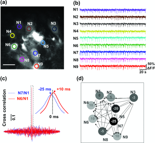
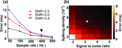
In the nervous system, most information is encoded in neural networks by dynamic patterns of activity.1 To characterize the spatiotemporal patterns of these neural networks represents a critical issue in modern neuroscience.2 Developed by Santiago Ramón y Cajal, the principles of specific connectivity and dynamic polarization purport that signals in a neural circuit travel only in one direction. This one-way flow of neuronal signaling was applied to all components of neural circuits, and gave rise to a logical set of rules to establish the correct flow of information and wiring of neural circuits.3 Currently, functional magnetic resonance imaging (fMRI) and electrophysiological recording, combined with analysis methods such as the Granger causality,4, 5, 6 symbolic transfer entropy,7 and Kullback–Leibler entropy,8 have proved to be helpful in the measurement of the brain activation for macrocircuits of both local- and whole-brain networks. These methods have been effectively applied in the identification of a neural network activation.9 It has been reported that microcircuits have an indispensable role in pursuing the mechanisms that underlie brain function, and the activation pattern of microcircuits have been successfully explored with cellular spatial resolution by using two-photon fluorescence microscopy.10, 11, 12 However, the limited data acquisition rate of the current two-photon microscopy (approximately several frames/s) is not fast enough for tracking the neural activities and signaling at the millisecond level;10 therefore, it remains difficult to examine the direction of activation and establish a directional cellular neuronal network. Here, we demonstrated the construction of a directional neural network with a cellular resolution using a fast two-photon microscopic imaging technique and cross correlation data analysis. A random access two-photon fluorescence microscope based on acousto-optical deflectors (AODs) was combined with calcium fluorescence labeling, enabling us to detect the calcium transients of synchronous firing in neural networks with a higher sampling rate. Thereby, millisecond scale activation delays among neurons in the population can be estimated from these calcium traces, and can be established based on the direction of network activation. A custom-built random access two-photon fluorescence microscope was applied to detect calcium fluorescence traces of neuron populations from rat brain slices. The design of this microscope was based on AOD, which delivers a laser beam to the preselected neurons of interest in an inertia-less manner.13, 14, 15, 16, 17 In the system, a mode locked laser of 800 nm was applied to excite calcium dyes, and two orthogonal AODs (DTS-XY, AA Opto-Electronic) were used for x–y deflection of the laser beam. A single dispersive prism is used and placed in a direction 45° relative to the x-axis to compensate both spatial and temporal dispersions.13 The lateral resolution of the system is 0.75 μm and the axial resolution of the system is 2.3 μm (40 × water immersion objective, N.A. = 0.8). The field of view is 220 μm × 220 μm. Hippocampus slices (350-μm thick) were obtained from postnatal 14 to 20 day wistar rats, and then labeled with acetoxymethyl ester (AM) calcium dyes Fura-2/AM, as previously reported.18 Briefly, slices were placed in 100 μl artificial cerebrosphinal fluid (ACSF) containing 40 μm Fura-2/AM, 0.005% Pluranic F-127, and 1% dimethylsulphoxide (DMSO). After labeling, the structural image of neural networks in the hippocampus CA1 region was captured by raster scans, and roughly 10 preselected regions of interest (ROIs) were imaged in a random-access scan pattern at a 0.4 kHz sampling rate, taking into account the balance between the sampling rate and the signal-to-noise ratio (SNR). The total effective dwell time was 250 μs/cell, and the sampling rate was 400 fps when 10 neurons were preselected as regions of interest in this experiment. 4-Aminopyridine, a bath-applied potassium channel blocker,19 was used at a final concentration of 100 μm to induce epileptiform discharging of the local neuron population. To establish the information flow and identify the neural circuit wiring, the action potential time which lags between two neurons from their cross correlation, must be determined. The calcium transients of neurons are triggered by action potentials, the calcium traces are approximately equal to the convolution of action potential sequence with the calcium trace template induced by a single action potential.10, 11 Therefore, the neural signal flow can be established directly from the neural calcium transients using these principles. By calculating the cross correlation between calcium traces x(t) and y(t) and the time corresponding to the maximum of the cross correlation sequence, the delay time τ between x(t) and y(t) can be determined. If τ is less than zero, the activation direction proceeds from x(t) to y(t) (expressed as symbol x→y). Otherwise, the directional activation is noted as y→x. Here, the calcium traces were filtered with a 0.01 Hz high-pass and 50 Hz low pass Butterworth filter to eliminate the influences of noises on the estimation precision of the delay time; it is essential to filter the calcium traces in view of neural activity frequency and calcium transient duration.20 The estimation precision of the delay time between calcium traces depends on the characters of the data, such as SNR, sampling frequency, and spike density. Here, SNR was defined as the ratio of the amplitude of calcium transients to the standard deviation of noise, and the spike density was defined as the ratio of the product of the calcium spike number and the duration of calcium transients during the recording period. To estimate the precision of the directional networks, we generated stimulation data as the raw calcium traces with the same data characters including SNR, sampling frequency, spike density, and noise level. The standard deviation of the delay time estimation was then calculated through cross correlation analysis of the stimulation data, which was used to evaluate the estimation precision of the real calcium traces. Through fast optical recording and cross correlation data analysis procedures, the direction of multicell network activation with cellular resolution was identified. Using the random access ability of the acousto-optic deflector, only the preselected regions were scanned. Thereby, the acquisition rate of the calcium signal from neurons of interest was increased without sacrificing the dwell time or the signal-to-noise ratio. Figure 1a shows a neural population in the rat hippocampal slice CA1 labeled with Fura-2/AM, and Fig. 1b displays simultaneous calcium transients of the neural population upon 100 μm 4-AP stimulation with a sampling rate of 400 Hz. Figure 1 establishes that the neuronal population fired calcium transients occur in a similar dynamic pattern. Cross correlations of calcium traces from neuron pairs were conducted, as shown in Fig. 1c, and the correlation coefficients ranged from 0.55 to 0.69. From the cross correlation sequence, the activation delay time of the neuron pairs N7/N1 and N6/N1 were established as −25 and +10 ms, respectively. Based on the delay time between each neuron pair, the directional connection and wiring of the neural circuits can be mapped as in Fig. 1d. In Fig. 1, the circles represent neurons, and the values depicted within these circles indicate the delay time relative to neuron N1. Additionally, the arrows between each neuron pair indicated the activation direction in the neural network. The results of several experimental trials from this neuron population demonstrated that the directionality of neural network activation was stationary, which validated the reliability of the directional networks that were constructed. Fig. 1Construction of a directional neural network of cellular resolution. (a) A cell population in rat hippocampal slice stained with Fura-2/AM. The circles indicate the preselected neurons. (b) Calcium transients of the nine individual neurons in epilepsy. The lines’ colors correspond to preselected neurons of the same color in (a), and fast calcium imaging reveals hypersynchronous neural discharge activity. (c) Cross correlation of neuron pairs N7/N1 and N6 /N1 with magnified fitted curves that show the delay time to be −25 and 10 ms, respectively. (d) Directional neural network topography. A topography of signal flow was mapped, and circles represent neurons in (a), and their darkness indicates the delay time (values are shown in the circles) of their calcium transients relative to neuron N1. Arrows between each two neurons indicate the direction of the neural network activation. Bar: 20 μm.  It should be noted that the precision of the calculated delay time between the neuron pair is actually dependent on the system performance and experimental conditions, such as sampling rate, SNR, and spike density. To evaluate the precision of this delay time, we generated simulation data with the same characters as the real calcium traces and calculated the error. First, we analyzed the real calcium traces to acquire the characters (i.e., SNR, spike density, sampling frequency, noise level, and calcium transient template). Based on these parameters, groups of 300 s long simulation data with different parameters were generated. These simulation data were then analyzed with the same cross correlation procedures to calculate the analysis error, as shown in Fig. 2. Figure 2a displays the analysis error of the delay time of the simulation data with a spike density of 0.05 (roughly 50 calcium spikes in the 300 s period) and SNR ranging from 2.5 to 3.5 at different sampling rates. Figure 2 demonstrated that a high sampling rate and high SNR is important for ensuring the precision of the delay time. In Fig. 2, the arrow indicates the error level, which is less than 2 ms, of the real calcium traces (400 Hz sampling rate) and an SNR of more than 3.0. Figure 2b shows the error of the delay time of simulation data at the 400 Hz sampling rate with different spike densities and SNR levels. The results indicated that increased SNR and spike density can improve the analysis precision. The star in Fig. 2 indicates the error level of the real calcium traces with spike density (0.05) and SNR (∼3.2), which ranges from 1.2 to 1.8 ms. Therefore, these results from the simulation data confirm the precision and reliability of the proposed fast optical recording and cross correlation analysis procedures. Fig. 2Time precision of the cross correlation procedures analyzed using simulation data. (a) The delay time error of simulation data with spike density (0.05), SNR (2.5 to 3.5) at different sampling rates. The arrow indicates the error level of the real calcium traces with 400 Hz sampling rate and SNR of more than 3.0. (b) The error at 400 Hz sampling rate with different spike densities and SNR levels. The star indicates the error level of the real calcium traces with spike density 0.05 and SNR about 3.2.  In the present study, the results demonstrated the construction of a directional neural network with high spatial and temporal resolution requires several essential elements including a multicell recording technique with a fast sampling rate and an adequate SNR level, suitable analysis method, and appropriate subjects. Two-photon calcium imaging is a powerful tool for monitoring the activity of multiple neurons in brain tissue.12 However, the sampling rate of a conventional system for recording calcium traces from multiple neurons is scarcely beyond 100 Hz without lowering the SNR. It remains difficult to identify neural population activities at a millisecond scale.10 In our study, we explored the random scanning mode and optimized the two-photon microscopy to increase the sampling rate. Because the signal-to-noise ratio of the calcium traces remain appropriate, the analysis precision of the calcium traces from the fast optical recording can be ensured,13, 14, 15 as shown in Figs. 2a, 2b. In addition, suitable analysis methods are also indispensable. For instance, the cross correlation algorithm is computationally simple and can be implemented as an efficient procedure for analysis. Finally, a synchronously firing network ensures the conformity of calcium traces from different neurons. The directional neural network contains information on not only the network connectivity, but also the activation direction. The construction of directional neural networks has spawned further research on the information processing mechanisms that are based on dynamic activation patterns.4, 5, 7, 9, 21 To date, the current directional neural networks focus mostly on the brain area level. Here, we constructed the directional neural network in local neural populations with a cellular resolution and a millisecond delay time precision. The cellular resolution directional network improves our knowledge by revealing the neurophysiological basis of the information processing and integration of brain microcircuits. In addition, the directional network can be an important indicator for the source of network oscillations.22, 23 Finally, the identification of some oscillations’ cellular pacemaker, such as the epileptogenic focus, will likely accelerate research on nervous system disorders in both basic and clinical research associated with the medical sciences. AcknowledgementsThis work was supported by the National Natural Science Foundation of China (Grant Nos. 30900331 and 61008053) and the National Science Fund for Distinguished Young Scholars (Grant No. 30925013). ReferencesW. Gobel and
F. Helmchen,
“In vivo calcium imaging of neural network function,”
Physiology, 22 358
–365
(2007). https://doi.org/10.1152/physiol.00032.2007 Google Scholar
R. Milo, S. Shen-Orr, S. Itzkovitz, N. Kashtan, D. Chklovskii, and
U. Alon,
“Network motifs: simple building blocks of complex networks,”
Science, 298 824
–827
(2002). https://doi.org/10.1126/science.298.5594.824 Google Scholar
J. DeFelipe,
“From the connectome to the synaptome: an epic love story,”
Science, 330 1198
–1201
(2010). https://doi.org/10.1126/science.1193378 Google Scholar
A. Brovelli, M. Ding, A. Ledberg, Y. Chen, R. Nakamura, and
S. L. Bressler,
“Beta oscillations in a large-scale sensorimotor cortical network: directional influences revealed by Granger causality,”
Proc. Natl. Acad. Sci. U.S.A., 101 9849
–9854
(2004). https://doi.org/10.1073/pnas.0308538101 Google Scholar
R. Goebel, A. Roebroeck, D. S. Kim, and
E. Formisano,
“Investigating directed cortical interactions in time-resolved fMRI data using vector autoregressive modeling and Granger causality mapping,”
Magn. Reson. Imaging, 21 1251
–1261
(2003). https://doi.org/10.1016/j.mri.2003.08.026 Google Scholar
M. Kaminski, M. Ding, W. A. Truccolo, and
S. L. Bressler,
“Evaluating causal relations in neural systems: granger causality, directed transfer function, and statistical assessment of significance,”
Biol. Cybern., 85 145
–157
(2001). https://doi.org/10.1007/s004220000235 Google Scholar
M. Staniek and
K. Lehnertz,
“Symbolic transfer entropy,”
Phys. Rev. Lett., 100 158101
(2008). https://doi.org/10.1103/PhysRevLett.100.158101 Google Scholar
C. J. Honey, R. Kotter, M. Breakspear, and
O. Sporns,
“Network structure of cerebral cortex shapes functional connectivity on multiple time scales,”
Proc. Natl. Acad. Sci. U.S.A., 104 10240
–10245
(2007). https://doi.org/10.1073/pnas.0701519104 Google Scholar
E. Bullmore and
O. Sporns,
“Complex brain networks: graph theoretical analysis of structural and functional systems,”
Nat. Rev. Neurosci., 10 186
–198
(2009). https://doi.org/10.1038/nrn2575 Google Scholar
B. F. Grewe, D. Langer, H. Kasper, B. M. Kampa, and
F. Helmchen,
“High-speed in vivo calcium imaging reveals neuronal network activity with near-millisecond precision,”
Nat. Methods, 7 399
–405
(2010). https://doi.org/10.1038/nmeth.1453 Google Scholar
E. Yaksi and
R. W. Friedrich,
“Reconstruction of firing rate changes across neuronal populations by temporally deconvolved Ca2+ imaging,”
Nat. Methods, 3 377
–383
(2006). https://doi.org/10.1038/nmeth874 Google Scholar
E. A. Mukamel, A. Nimmerjahn, and
M. J. Schnitzer,
“Automated analysis of cellular signals from large-scale calcium imaging data,”
Neuron., 63 747
–760
(2009). https://doi.org/10.1016/j.neuron.2009.08.009 Google Scholar
S. Zeng, X. Lv, C. Zhan, W. R. Chen, W. Xiong, S. L. Jacques, and
Q. Luo,
“Simultaneous compensation for spatial and temporal dispersion of acousto-optical deflectors for two-dimensional scanning with a single prism,”
Opt. Lett., 31 1091
–1093
(2006). https://doi.org/10.1364/OL.31.001091 Google Scholar
X. Lv, C. Zhan, S. Zeng, W. R. Chen, and
Q. Luo,
“Construction of multiphoton laser scanning microscope based on dual-axis acousto-optic deflector,”
Rev. Sci. Instrum., 77 046101
(2006). https://doi.org/10.1063/1.2190047 Google Scholar
Y. Wu, X. Liu, W. Zhou, X. Lv, and
S. Zeng,
“Observing neuronal activities with random access two-photon microscope,”
J. Innovative Opt. Health Sciences, 2 67
–71
(2009). https://doi.org/10.1142/S179354580900036X Google Scholar
G. D. Reddy, K. Kelleher, R. Fink, and
P. Saggau,
“Three-dimensional random access multiphoton microscopy for functional imaging of neuronal activity,”
Nat. Neurosci., 11 713
–720
(2008). https://doi.org/10.1038/nn.2116 Google Scholar
V. Iyer, T. M. Hoogland, and
P. Saggau,
“Fast functional imaging of single neurons using random-access multiphoton (RAMP) microscopy,”
J. Neurophysiol., 95 535
–545
(2006). https://doi.org/10.1152/jn.00865.2005 Google Scholar
D. Smetters, A. Majewska, and
R. Yuste,
“Detecting action potentials in neuronal populations with calcium imaging,”
Methods, 18 215
–221
(1999). https://doi.org/10.1006/meth.1999.0774 Google Scholar
H. V. Srinivas,
“Epilepsy: The future scenario,”
Ann. Indian Acad. Neurol., 13 2
–5
(2010). https://doi.org/10.4103/0972-2327.61269 Google Scholar
M. Bekara and
M. van der Bann,
“A new parametric method for time delay estimation,”
IEEE Trans. Acoust., Speech, Signal Process. III, 3 1033
–1036
(2007). Google Scholar
P. Fries,
“A mechanism for cognitive dynamics: neuronal communication through neuronal coherence,”
Trends Cogn. Sci., 9 474
–480
(2005). https://doi.org/10.1016/j.tics.2005.08.011 Google Scholar
M. J. Morrell,
“Working toward an epilepsy cure,”
Curr. Neurol. Neurosci. Rep., 3 323
–324
(2003). https://doi.org/10.1007/s11910-003-0009-x Google Scholar
P. H. Manninen, S. J. Burke, R. Wennberg, A. M. Lozano, and
H. El Beheiry,
“Intraoperative localization of an epileptogenic focus with alfentanil and fentanyl,”
Anesth. Analg., 88 1101
–1106
(1999). https://doi.org/10.1097/00000539-199905000-00025 Google Scholar
|

