
|
|
1.IntroductionThe human sclera is a turbid, nontransparent medium covering about 80% of the eye ball and serving as a protective membrane. It is a strong fibrous tissue which mainly consists of conjunctive collagen fibers forming parallel bundles of different diameters that are organized in irregular interwoven layers.1, 2 The human sclera is composed mainly of type-I collagen fibrils (50% to 70%), which mostly distribute among the equator and posterior pole region of the eyeball. Collagen types III, V, and VI have also been found in the sclera.3, 4 Myopia is a common ocular disability throughout the world. Myopia affects up to 30% of the general population in the USA and Europe, and up to 60% of Asians.5, 6, 7, 8, 9 High myopia is characterized by sclera thinning and localized ectasia of the posterior sclera. The progression of scleral thinning can be attributed to an interrupted emmetropization mechanism after visual deprivation, such as a cataract, corneal opacity, or a shift in the focal plane by negative lenses.10 On the other hand, the condition may be due to genetic problems, such as a Marfan syndrome (elastin defect), Ehler-danros syndrome (collagen type-I and -III), Osteogenesis imperfecta (collagen type-I), and Pseudozanthoma elasaticum (elastin defect) with a lack of intermolecular cross-links of collagen or elastin.11, 12 To reduce myopic progression, various clinical trials such as pharmaceutical agents, progressive addition lenses, rigid gas-permeable contact lenses, orthokeratotic lens, and scleral reinforcement operations, have yielded negative or positive results. 13, 14, 15, 16, 17, 18, 19 Recently, some studies reported that the cross-linking induced by the photosensitizer riboflavin and ultraviolet A (UVA) irradiation of 370 nm lead to a significant increase in the biomechanical rigidity efficiency of scleral collagens, which can prevent myopic progression.20, 21 However, they provided no information on the structural changes and chemical bonds in the biomolecule interactions of the scleral collagen. Raman spectroscopy has many advantages when analyzing biological tissues, such as less water interference, nondestructive analysis, and high sensitivity. Furthermore, it can provide information on the structure and interactions of biomolecules in their microenvironment within intact cells and tissues. This method was used successfully to examine the mechanism of glutaraldehyde or dimethyl suberimidate cross-linking in pericardium tissue.22, 23 Atomic force microscopy (AFM) is a promising tool for observing the surface topography or nanostructure of biological tissues because it provides high spatial resolution on the nanometer scales as well as the three-dimensional ultrastructure of the surface morphology.24, 25 Therefore, this study investigated the effect of cross-linking with riboflavin-UVA irradiation on the chemical bonds and ultrastructure of human sclera tissues using Raman spectroscopy, AFM, and histology. 2.Material and Methods2.1.Sample PreparationSclera from a human donor eye (age 70 years, male) was obtained from the Eye Bank of Kyung Hee University Medical Center, Seoul, Korea. The donor sclera had negative serologic tests for hepatitis, syphilis, and human immune deficiency virus. The eye was prepared by surgically removing the internal ocular structures to leave only the scleral shell. The human sclera tissue was preserved in 75% ethanol. 2.2.Cross-Linking ProcedureA 0.1% riboflavin photosensitizer solution (3 mg riboflavin-5-phosphate in 3 ml 20% dextran 500) was instilled into the prepared scleral tissues for 10 min before UVA irradiation. UVA irradiation (370 nm) was applied using UVA light source (UV-XTM, IROC AG, Zürich Switzerland) with an irradiance of 3 mW/cm2 at a distance of 4 cm from the sclera for 30 min. 2.3.Raman SpectroscopyRaman spectroscopy (InVia Raman, Renishaw, United Kingdom) was performed to characterize the chemical composition and molecular structure of normal and cross-linked sclera tissues. A λ = 785 nm with a power of 200 mW was used as the excitation source. The spectra were recorded by scanning the 200 to 3200 cm−1 region with an acquisition time of 10 s for each scan. 2.4.AFM MeasurementThe sclera specimens were dehydrated in a graded series (60%, 70%, 80%, 90%, 95%, and 100% ethanol with 3 changes) of ethanol and dried in air. The sclera surface was examined by AFM (NANOS N8 NEOS, Bruker, Herzogenrath, Germany), which was operated in noncontact mode (nominal spring constant 0.2 N/m) in ambient air at room temperature. The topography was obtained using a silicon cantilever with an integral pyramidal shaped tip (SICONG, Santa Clara, California). The AFM images had an image resolution of 512 × 512 pixels, and a scan speed was of 1.0 line/s. A scanning probe image processor (SPIP version 4.8, Image Metrology, Lyngby Denmark) was used to analyze and compare the morphology of the normal and cross-linked scleral tissues. 2.5.Histological AnalysisBoth normal and cross-linked sclera tissues were dehydrated in a graded series of ethanol followed by xylene (70% v/v ethanol for 2 h, 100% v/v ethanol for 2 h, and xylene for 1 h) and embedded in paraffin at 60°C for 2 h. Sections (4-μm thick) from each block were mounted on a glass microscope slide. After removing the embedding medium, the sectioned sclera tissue was stained with Masson's trichrome stain. The stained slices were observed by optical microscopy (ScanScope CS, APERIO, California). 3.ResultsFigure 1 presents the Raman spectra of normal and cross-linked human sclera tissues in the range of (a) 200 to 3200 cm−1, (b) 200 to 1800 cm−1, and (c) 2800 to 3200 cm−1. Table 1 summarizes the Raman shift and the assignment of the bands, which are discussed in terms of riboflavin-UVA and collagen interactions.26, 27, 28, 29, 30, 31 Fig. 1Raman spectra of the normal and cross-linked human sclera tissue in three spectral ranges of (a) 200 to 3200 cm−1, (b) 200 to 1800 cm−1, and (c) 2800 to 3200 cm−1, respectively. The peak assignments are given in Table 1. Raman peaks, which are discussed in terms of the cross-linking, are in the dashed line. 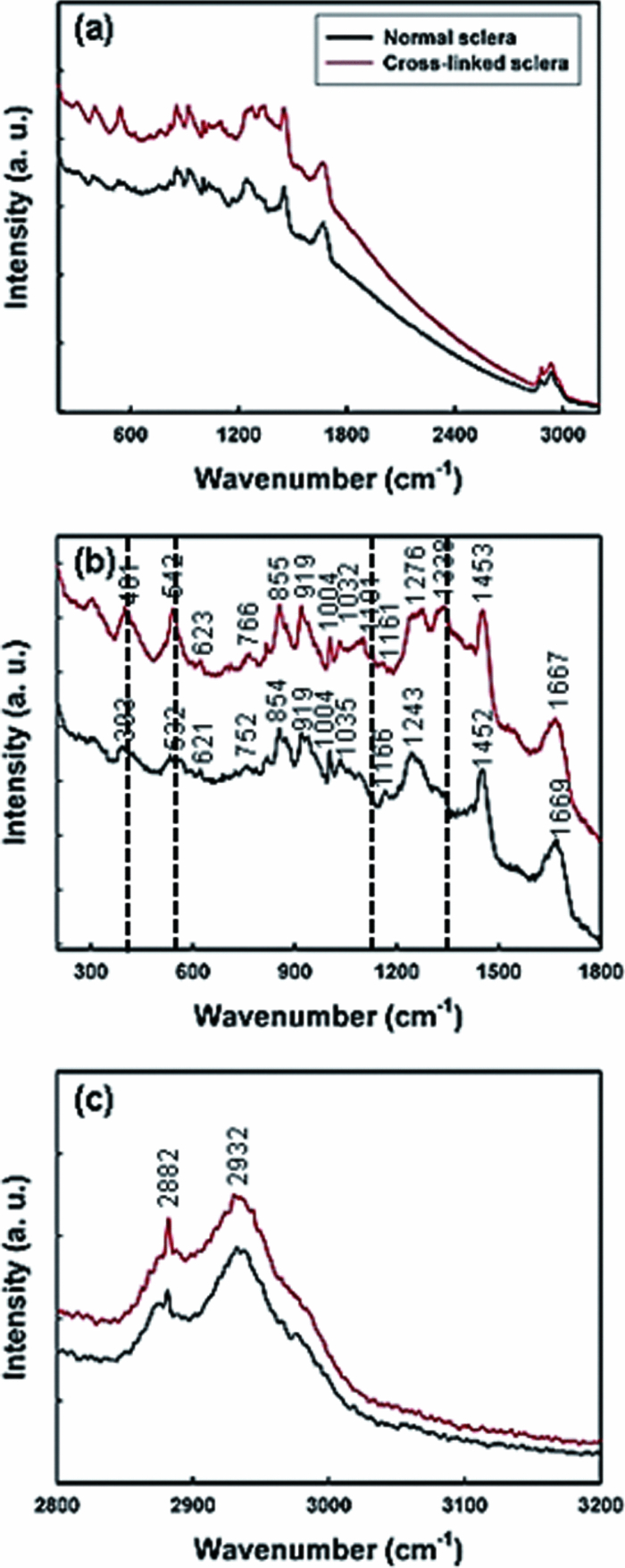 Table 1Band assignments for the Raman spectra of human sclera in normal and cross-linking (Refs. 26, 27, 28, 29, 30, 31).
There was a strong dependency between the intensity of certain Raman bands at the amide bands of 1276 and 1668 cm−1. The Raman bands at 855 and 919 cm−1 were assigned to the C-C stretching vibration of the proline ring in collagen. The band at 1004 cm−1 (C-C aromatic ring), which is typical for the collagen, was observed. The methyl (CH3) and methylene (CH2) deformation vibration mode at 1452 cm−1 also features the Raman scattering in collagen. The scleral tissue treated by riboflavin-UVA showed not only a shift in the 393 cm−1 (CCC) stretch band toward 401 cm−1 and a simultaneous shift in the 532 cm−1 (S-S) stretch band toward 542 cm−1, but also more intense and sharp Raman peaks. The broad peak at approximately 1322 to 1338 cm−1 was assigned to CH2 deformation. In Fig. 1c, the peak at 2882 cm−1 was attributed to the (CH2) antisymmetric stretching mode, and a broad spectra peak at 2932 cm−1 was assigned to the (CH3, CH2) stretching mode. Figure 2 shows the surface topography, three-dimensional images, and line profiles of normal [Figs. 2a, 2b, 2c] and cross-linked [Figs. 2d, 2e, 2f] human scleral tissues. For the normal sclera specimen, the collagen fibrils showed a regular parallel arrangement with a clear axial, and a regular transverse D-periodic banding pattern consisting of high ridges and shallow grooves was distinctly seen; whereas the spatial pattern of fibrils within cross-linked sclera tissue was tangled and running in different directions. Fig. 2AFM topography (a) and three-dimensional image (b) for a normal human scleral tissue. The curve (c) shows the line profile along line 1 for the collagen fibril. AFM topography (d) and three-dimensional image (e) for cross-linked human scleral tissue. The curve (f) shows the line profile along line 2 for the cross-linked collagen fibril. The spatial pattern of fibrils within cross-linked sclera tissue was tangled and running in different directions (arrowheads).  Figures 3a, 3b show the Masson's trichrome staining of the normal and cross-linked sclera tissues (magnification × 400). The difference in stained collagen as a blue color was clearly identified between the normal and cross-linked specimens. The cross-linked scleral tissue showed a dense collagen bundle compared to the normal sclera tissues. Fig. 3Histology section of normal (a) and cross-linked (b) human sclera tissues, stained with Masson's trichrome (magnification × 400), where collagen stains blue, nuclei stain black, and cell bodies stain red. The cross-linked scleral tissue showed a dense collagen bundle compared to the normal sclera tissues.  4.DiscussionThe sample preservation of this study was based on the clinical preservation method, used ophthalmologically in our hospital. This method may affect the structure of scleral tissues, but since this study examined and compared the difference between the normal and cross-linked sclera tissues using the same method, that effect may not be a problem. Cross-linking is a minimally invasive procedure in which a chemical agent is applied to the residual cornea or sclera after epithelium removal. This chemical agent initiates the formation of new molecular bonds between the collagen fibrils and lamellae, either by itself or when exposed to UV light. New bonds would be the most likely to increase the mechanical strength of the cornea or sclera because they could physically link individual collagen fibrils, and entire adjacent lamellae of the cornea or sclera stroma. 20, 21, 32, 33, 34, 35, 36 Clinical collagen cross-linking of keratoconus corneas induced by the photosensitizer riboflavin and UVA irradiation of 370 nm prevents further corneal stroma thinning associated with keratoconus by increasing corneal rigidity.32, 33, 34, 35 Wollensak reported that the cross-linking induced by the riboflavin-UVA irradiation leads to a significant increase in the biomechanical rigidity efficiency of scleral collagens, which may prevent myopic progression.20, 21 However, they provided no information on the structural changes and chemical bonds in the biomolecule interactions of the collagen. Human scleral tissue contains approximately 50% collagen by weight, consisting mainly of type-I collagen.37, 38 Generally, collagens are identified as proteins consisting of three polypeptide chains, assembled with triple-helical domains and containing (Gly-X-Y)n amino acid repeat sequences, where X and Y are often proline and hydroxyproline, respectively.38 In this study, the Raman spectra of human sclera tissue are consistent and feature the characteristic collagen bands.26, 31 The amide I (1655 to 1667 cm−1) and amide III (1241 to 1276 cm−1) bands, which represent peptide bonds within the proteins and in the collagen, indicate the stabilization of a subfibrillar triple helical structure by the formation of interchain hydrogen bonds between the N-H groups of glycines and the C=O groups of prolines in the neighboring chains.39, 40 The amide I band corresponds to the peptide carbonyl stretching vibration, whereas the amide III band originates from the NH in plane deformation at 1276 cm−1 coupled to the CN stretching mode at 1243 cm−1 (Table 1). In addition, a strong C-C stretch band around 855 and 919 cm−1 supports the presence of the helical conformation in a collagen molecule.26 The interactions of the riboflavin-UVA with the collagen from human sclera result in formation of stable cross-links, which the number of cross-links in collagen increases. McCall explained the mechanism of cross-linking with riboflavin-UVA in the corneal tissue, which the riboflavin-sensitized UVA photoreaction generates free radicals and so-called reactive oxygen species, like superoxide anion (O2 −), hydroxyl radical (•OH), or hydrogen peroxide (H2O2) mainly via the so-called type-I pathway of photosensitized oxidation.36 This chemical modification induces changes in the Raman spectra of the sclera tissues. The analysis of the position and the intensity of the Raman bands for normal and cross-linked sclera tissues allowed us to recognize the following types of the riboflavin-UVA and collagen interactions. When the scleral tissue was treated by riboflavin-UVA, it can observe not only a shift of the 393 cm−1 (CCC) stretch band toward 401 cm−1 and a simultaneous shift in the 532 cm−1 (S-S) stretch band toward 542 cm−1, but also more intense and sharp Raman peaks. A weak band around 1126 cm−1 (CN stretch) appeared which could be explained in terms of C-N bond formation by riboflavin-UVA. Furthermore, cross-linked scleral tissue showed an increase in the band intensity around 1322 to 1338 cm−1, which is assigned to the CH2 deformation and would be attributed to a relative increase of the CH2 chains according to the cross-linking effect. Therefore, Raman spectroscopy can identify the changes in molecular structure and chemical composition of collagen fibrils in both normal and cross-linked human scleral tissues. AFM is being widely applied to study surface topography of many biological structures, at high resolution. The ultrastructure of corneal collagen fibrils as well as scleral collagen fibrils was first studied using AFM by Fullwood 41 They reported that cross-bridge structures were sometimes visible connecting the collagen fibrils in both cornea and sclera. Meller also reported quantitative measurements of individual collagen fibrils in human cornea and sclera.42 In the present study, the surface topography of normal sclera tissue is consistent with that previously reported, in which regular transverse D-periodic banding pattern of approximately 67 ± 6 nm.41 The sclera tissues with riboflavin-UVA treatment showed the tangled and cross-bridge nanostructures with a swelling effect and a running in different directions each other. It is likely that this change is responsible for formation of cross-links between collagen molecules within collagen fibrils. To investigate the cross-linking effect of the human sclera, histological analysis of normal and cross-linked sclera tissues was performed using Masson's trichrome staining method to visualize the extracellular matrix components, particularly the collagen fibrils in the sclera. A dense collagen bundle in the crossed-linked sclera tissue might be caused by the cross-linking effect by the riboflavin-UVA treatment. In this study, there are some limitations including the number of samples and the effects with age and investigating regions in sclera. Therefore, further studies will be necessary to determine the relevant clinical implications of the findings. The is further dictated to obtain a reliable result by means of more sample size and to reveal the relationship between the age and regions in sclera. In summary, this study showed that human scleral collagen can be cross-linked effectively using riboflavin-UVA irradiation, leading to a significant increase in the collagen bundle. Raman spectroscopy and AFM were used to examine the structural changes and chemical bonds in cross-linked human sclera tissues by riboflavin-UVA irradiation. In addition, the scleral tissues were examined histologically by optical microscopy to evaluate the cross-linking effect. The Raman spectra of the normal and cross-linked human sclera tissues revealed different types of the riboflavin-UVA and collagen interactions, which could be identified from their unique peaks, intensity, and shape. Therefore, Raman spectroscopy can prove to be a powerful tool for examining chemical bonds in collagenous tissues at the molecular level. After sclera collagen cross-linking, AFM image revealed interlocking arrangements of collagen fibrils. The observed changes in the surface topography of the collagen fibrils, as well as in their chemical bonds in the tissue, support the formation of interfibrilar cross-links in sclera tissues. AcknowledgmentsThis study was supported by the Industrial Core Technology Development Program funded by the Ministry of Knowledge Economy (No. 10037379, Development of Multi X-ray Source and Tomosynthesis System based on Nano Materials). This study was also supported by a 2010 Research Grant from Kangwon National University. ReferencesY. Komai and
T. Ushiki,
“The three-dimensional organization of collagen fibrils in the human cornea and sclera,”
Invest. Ophthalmol. Vis. Sci., 32
(8), 2244
–2258
(1991). Google Scholar
J. A. Summers Rada,
S. Shelton, and
T. T. Norton,
“The sclera and myopia,”
Exp. Eye Res., 82
(2), 185
–200
(2006). https://doi.org/10.1016/j.exer.2005.08.009 Google Scholar
F. W. Keeley,
J. D. Morin, and
S. Vesely,
“Characterization of collagen from normal human sclera,”
Exp. Eye Res., 39
(5), 533
–542
(1984). https://doi.org/10.1016/0014-4835(84)90053-8 Google Scholar
P. G. Watson and
R. D. Young,
“Sclera structure, organization and disease. A review,”
Exp. Eye Res., 78
(3), 609
–623
(2004). https://doi.org/10.1016/S0014-4835(03)00212-4 Google Scholar
N. A. McBrien and
A. Gentle,
“Role of the sclera in the development and pathological complications of myopia,”
Prog. Retin Eye Res., 22
(3), 307
–338
(2003). https://doi.org/10.1016/S1350-9462(02)00063-0 Google Scholar
L. L. Lin,
Y. F. Shih,
C. K. Hsiao, and
C. J. Chen,
“Prevalence of myopia in Taiwanese schoolchildren: 1983 to 2000,”
Ann. Acad. Med. Singapore, 33
(1), 27
–33
(2004). Google Scholar
S. –M. Saw,
E. C. Shih-Yen,
A. Koh, and
D. Tan,
“Interventions to retard myopia progression in children; an evidence-based update,”
Ophthalmology, 109
(3), 415
–421
(2002). https://doi.org/10.1016/S0161-6420(01)00972-1 Google Scholar
S. M. Saw,
H. M. Wu,
B. Seet,
T. Y. Wong,
E. Yap,
K. S Chia,
R. A. Stone, and
L. Lee,
“Academic achievement, close up work parameters, and myopia in Singapore military conscripts,”
Br. J. Ophthalmol., 85
(7), 855
–860
(2001). https://doi.org/10.1136/bjo.85.7.855 Google Scholar
I. Morgan and
K. Rose,
“How genetic is school myopia,”
Prog. Retin. Eye Res., 24
(1), 1
–38
(2005). https://doi.org/10.1016/j.preteyeres.2004.06.004 Google Scholar
J. A. Summers Rada,
S. Shelton, and
T. T. Norton,
“The sclera and myopia,”
Exp. Eye Res., 82
(2), 185
–200
(2006). https://doi.org/10.1016/j.exer.2005.08.009 Google Scholar
G. Mechanic,
“Crosslinking of collagen in a heritable disorder of connective tissue: Ehlers-Danlos syndrome,”
Biochem. Biophys. Res. Commun., 47
(1), 267
–272
(1972). https://doi.org/10.1016/S0006-291X(72)80038-X Google Scholar
G. R. Bell,
“Biomechanical considerations of high myopia: part I – physiological characteristics,”
J. Am. Optom. Assoc., 64
(5), 332
–338
(1993). Google Scholar
J. Gwiazda,
L. Hyman,
M. Hussein,
D. Everett,
T. T Norton,
D. Kurtz,
M. C. Leske,
R. Manny,
W. M Tootle,
M. Scheiman, COMET Group,
“A randomized clinical trial of progressive addition lenses versus single vision lenses on the progression of myopia in children,”
Invest. Ophthalmol. Vis. Sci., 44
(4), 1492
–1500
(2003). https://doi.org/10.1167/iovs.02-0816 Google Scholar
M. H. Edwards,
R. W. Li,
C. S. Lam,
J. K. Lew, and
B. S. Yu,
“The Hong Kong Progressive Lens Myopia Control Study: study design and main findings,”
Invest. Ophthalmol. Vis. Sci., 43
(9), 2852
–2858
(2002). Google Scholar
J. Katz,
O. D. Schein,
B. Levy,
T. Cruiscullo,
S. M. Saw,
U. Rajan,
T. K. Chan,
C. Y. Khoo, and
S. J. Chew,
“A randomized trial of rigid gas permeable contact lenses to reduce progression of children's myopia,”
Am. J. Ophthalmol., 136
(1), 82
–90
(2003). https://doi.org/10.1016/S0002-9394(03)00106-5 Google Scholar
R. M. Siatkowski,
S. A. Cotter,
J. M Miller,
C. A. Scher,
R. S Crockett, and
G. D. Novack,
“Safety and efficacy of 2% pirenzepine ophthalmic gel in children with myopia: a 1-year, multicenter, double-masked, placebocontrolled parallel study,”
Arch. Ophthalmol., 122
(11), 1667
–1674
(2004). https://doi.org/10.1001/archopht.122.11.1667 Google Scholar
R. M. Siatkowski,
S. A. Cotter,
R. S. Crockett,
J. M. Miller,
G. D. Novack, and
K. Zadnik,
“Two-year multicenter, randomized, double-masked, placebo-controlled, parallel safety and efficacy study of 2% pirenzepine ophthalmic gel in children with myopia,”
J. AAPOS, 12
(4), 332
–339
(2008). https://doi.org/10.1016/j.jaapos.2007.10.014 Google Scholar
E. S. Avetisov,
E. P. Tarutta,
E. N. Iomdina,
M. I. Vinetskaya, and
L. D. Andreyeva,
“Nonsurgical and surgical methods of sclera reinforcement in progressive myopia,”
Acta Ophthalmol. Scand., 75
(6), 618
–623
(1997). https://doi.org/10.1111/j.1600-0420.1997.tb00617.x Google Scholar
A. A. Snyder and
F. B. Thompson,
“A simplified technique for surgical treatment of degenerative myopia,”
Am. J. Ophthalmol., 74
(2), 273
–277
(1972). Google Scholar
G. Wollensak and
E. Spoerl,
“Collagen crosslinking of human and porcine sclera,”
J. Cataract Refract. Surg., 30
(3), 689
–695
(2004). https://doi.org/10.1016/j.jcrs.2003.11.032 Google Scholar
G. Wollensak,
E. Iomdina,
D.-D. Dittert,
O. Salamatina, and
G. Stoltenburg,
“Cross-linking of scleral collagen in the rabbit using riboflavin and UVA,”
Acta Ophthalmol., 83
(4), 477
–482
(2005). https://doi.org/10.1111/j.1600-0420.2005.00447.x Google Scholar
M. Jastrzebska,
R. Wrzalik,
A. Kocot,
J. Z. Rejdak, and
B. Cwalina,
“Raman spectroscopic study of glutaraldehyde-stabilized collagen and pericardium tissue,”
J. Biomater. Sci. Polym. Ed., 14
(2), 185
–197
(2003). https://doi.org/10.1163/156856203321142605 Google Scholar
M. Jastrzebska,
J. Z. Rejdak,
R. Wrzalik,
A. Kocot,
B. Barwiński,
I. Mróz, and
B. Cwalina,
“Dimethyl suberimidate cross-linked pericardium tissue: Raman spectroscopic and atomic force microscopy investigations,”
J. Mol. Struct., 744–747
(3), 789
–795
(2005). https://doi.org/10.1016/j.molstruc.2004.11.040 Google Scholar
D. Meller,
K. Peters, and
K. Meller,
“Human cornea and sclera studied by atomic force microscopy,”
Cell Tissue Res., 288
(1), 111
–118
(1997). https://doi.org/10.1007/s004410050798 Google Scholar
S. Yamamoto,
H. Hashizume,
J. Hitomi,
M. Shigeno,
S. Sawaguchi,
H. Abe, and
T. Ushiki,
“The subfibrillar arrangement of corneal and scleral collagen fibrils as revealed by scanning electron and atomic force microscopy,”
Arch. Histol. Cytol., 63
(2), 127
–135
(2000). https://doi.org/10.1679/aohc.63.127 Google Scholar
B. G. Frushour and
J. L. Koening,
“Raman scattering of collagen, gelatin and elastin,”
Biopolymers, 14
(2), 379
–391
(1975). https://doi.org/10.1002/bip.1975.360140211 Google Scholar
H. G. M. Edwards,
D. W. Farwell,
J. M. Holder, and
E. E. Lawson,
“Fourier-transform Raman spectroscopy of ivory: II. Spectroscopic analysis and assignments,”
J. Mol. Struct., 435
(1), 49
–58
(1997). https://doi.org/10.1016/S0022-2860(97)00122-1 Google Scholar
X. Du and
Y. Liang,
“FT-Raman and FTIR spectroscopic studies of N-octadecanoyl-l-alanine amphiphiles,”
Spectrochim. Acta, Part A, 60
(1–2), 401
–404
(2004). https://doi.org/10.1016/S1386-1425(03)00238-5 Google Scholar
J. K. Tait,
H. G. Edwards,
D. W. Farwell, and
J. Yarwoodm,
“Fourier transform Raman spectroscopic examination of two amine-based epoxy resin crosslinking agents,”
Spectrochim. Acta, Part A, 51
(12), 2101
–2106
(1995). https://doi.org/10.1016/0584-8539(95)01456-5 Google Scholar
D. Lin-Vien,
N. B. Colthup,
W. G. Fateky, and
I. G. Grasselil, The Handbook of Infrared and Raman Characteristic Frequencies of Organic Molecules, Academic Press Ltd., London
(1991). Google Scholar
M. Janko,
P. Davydovskaya,
M. Bauer,
A. Zink, and
R. W. Stark,
“Anisotropic Raman scattering in collagen bundles,”
Opt. Lett., 35
(16), 2765
–2767
(2010). https://doi.org/10.1364/OL.35.002765 Google Scholar
C. Wittig-Silva,
M. Whiting,
E. Lamoureux,
R. G. Lindsay,
L. J. Sullivan, and
G. R. Snibson,
“A randomized controlled trial of corneal collagen cross-linking in progressive keratoconus: preliminary results,”
J. Refract. Surg., 24
(7), 720
–725
(2008). Google Scholar
F. Raiskup-Wolf,
A. Hoyer,
E. Spoerl, and
L. E. Pillunat,
“Collagen crosslinking with riboflavin and ultraviolet-A light in keratoconus: long-term results,”
J. Cataract Refract. Surg., 34
(5), 796
–801
(2008). https://doi.org/10.1016/j.jcrs.2007.12.039 Google Scholar
E. Spoerl,
M. Huhle, and
T. Seiler,
“Induction of cross-links in corneal tissue,”
Exp. Eye Res., 66
(1), 97
–103
(1998). https://doi.org/10.1006/exer.1997.0410 Google Scholar
G. Wollensak and
B. Redl,
“Gel electrophoretic analysis of corneal collagen after photodynamic cross-linking treatment,”
Cornea, 27
(3), 353
–356
(2008). https://doi.org/10.1097/ICO.0b013e31815cf66a Google Scholar
A. S. McCall,
S. Kraft,
H. F. Edelbauser,
G. W. Kidder,
R. R. Lundquist,
H. E. Bradsbaw,
Z. Dedeic,
M. J. C. Dionne,
E. M. Clement, and
G. W. Conrad,
“Mechanisms of corneal tissue cross-linking in response to treatment with topical riboflavin and long-wavelength ultraviolet radiation (UVA),”
Invest. Ophthalmol. Vis. Sci., 51
(1), 129
–138
(2010). https://doi.org/10.1167/iovs.09-3738 Google Scholar
F. W. Keeley,
J. D. Morin, and
S. Vesely,
“Characterization of collagen from normal human sclera,”
Exp. Eye. Res., 39
(5), 533
–542
(1984). https://doi.org/10.1016/0014-4835(84)90053-8 Google Scholar
P. G. Watson and
R. D. Young,
“Scleral structure, organization and disease,”
Exp. Eye. Res., 78
(3), 609
–623
(2004). https://doi.org/10.1016/S0014-4835(03)00212-4 Google Scholar
R. D. B. Fraser,
T. P. MacRae, and
E. Suzuki,
“Chain conformation in the collagen molecule,”
J. Mol. Biol., 129
(3), 463
–481
(1979). https://doi.org/10.1016/0022-2836(79)90507-2 Google Scholar
A. Rich and
F. H. C. Crick,
“The structure of collagen,”
Nature., 176
(4489), 915
–916
(1995). https://doi.org/10.1038/176915a0 Google Scholar
N. J. Fullwood,
A. Hammiche,
H. M. Pollock,
D. J. Hourston, and
M. Song,
“Atomic force microscopy of the cornea and sclera,”
Current Eye Res., 14
(7), 529
–535
(1995). https://doi.org/10.3109/02713689508998399 Google Scholar
D. Meller,
K. Peters, and
K. Meller,
“Human cornea and sclera studied by atomic force microscopy,”
Cell Tissue Res., 288
(1), 111
–118
(1997). https://doi.org/10.1007/s004410050798 Google Scholar
|


