

|
|
|
Diffuse reflectance spectroscopy has been explored to characterize tissue condition noninvasively in recent years.1,2 Diffuse reflectance spectra have been used to estimate the optical properties of tissues,3 which can be correlated with several important biophysical and biochemical parameters, such as hemoglobin information,2 tissue oxygenation,2 and average nuclei size4 in tissues for disease diagnoses. Nonetheless, one significant disadvantage of traditional diffuse reflectance spectroscopy is slow data acquisition when diffuse reflectance spectra at multiple locations are required due to the employment of fiber-optic probes, which can only perform optical measurements point by point. Multi-spectral imaging has been used to acquire diffuse reflectance images at multiple wavelengths.5 Even so, image acquisition can still be slow when the required spectral resolution is high. Although there have been efforts in the development of snapshot spectroscopic imaging techniques,6 the advantage of these techniques in quick image acquisition is usually compromised by slow post-processing, especially when the data dimension is large. Therefore, it is critical to develop a rapid spectral imaging technique, which could offer real-time acquisition of diffuse reflectance spectra for tissue measurements in multiple locations without sacrificing the spectral resolution. The reconstruction of diffuse reflectance spectra from color values taken by a color camera is a potential solution to this problem. In this approach, the color images of a large tissue area under white-light illumination are captured in real time by a color camera, which typically includes the color values in red, green, and blue bands at each pixel. Each color band covers a relatively wide range of wavelengths; thus, the signal is typically strong for fast imaging. Several methods, including pseudo-inverse,7 finite-dimensional modeling8 and Wiener estimation,9 have been explored to recover diffuse reflectance spectra from color values in multiple bands. Among them, Wiener estimation is one of the most frequently used methods because of its time efficiency.10 Even so, one serious problem of using this estimation method is that more than three color bands are usually necessary for sufficient accuracy,9 because of the underdetermined nature of the problem, in which three values corresponding to RGB colors are mapped to diffuse reflectance intensities at tens of or a few hundred wavelengths in a spectrum. As a result of inadequate Wiener estimation, the error in the estimated diffuse reflectance spectra could propagate and lead to larger errors in estimated optical properties11 and subsequently in estimated tissue parameters for diagnosis. Therefore, a fast method for the accurate estimation of diffuse reflectance spectra is needed in biomedical applications. In this paper, we propose a modified Wiener estimation method to address this problem by synthesizing new colors from the system matrix based on a calibration data set, which will provide additional information required for accurate Wiener estimation. The method has demonstrated significant improvement over traditional Wiener estimation in the estimation of diffuse reflectance spectra from color measurements acquired in vivo from human skin. This method consists of two stages and one post-processing step, i.e., the calibration stage, the test stage, and the selection step. In the calibration stage, as shown in Fig. 1, we first calculate the system matrix and initial Wiener matrix using the calibration data, which contain both measured RGB color values and the corresponding measured diffuse reflectance spectra. The estimation of the system matrix is an overdetermined problem and thus should be accurate because a diffuse reflectance spectrum contains many more data points than RGB values. Diffuse reflectance spectra can be estimated from measured RGB color values by using the initial Wiener matrix. Then, by applying a synthetic absorption filter, which contains three absorption bands different from RGB filters, to the system matrix, a modified system matrix can be created, which is used to generate two separate sets of three new color values from the measured and estimated diffuse reflectance spectra. The transmission spectra of the three synthetic absorption filters were determined by Gaussian functions with different center wavelengths: 500, 550, and 600 nm, and a standard deviation of 100 nm. The center wavelengths were selected to deviate from those of RGB filters in order to cover different information. Because of the high accuracy of the system matrix, the new color values estimated from the measured diffuse reflectance spectrum should be accurate and thus could be seen as the reference new color values. The new color values generated from the estimated diffuse reflectance spectra are less accurate, which will be called estimated new color values to facilitate the discussion next. The relation between the estimated new color values and the reference new color values can be found by using the following two strategies: 1. to model each reference new color value as a second-order polynomial function of estimated new color values; 2. to record the differences in each color value between two sets. Moreover, the reference new color values combined with the original RGB values are used to create a modified Wiener matrix for the second-round estimation of diffuse reflectance spectra in the test stage, which will be more accurate than using the RGB values alone because of the increase in the number of available color bands. In the test stage, as shown in Fig. 2, the initial estimated diffuse reflectance spectrum will be computed by applying the initial Wiener matrix to measured RGB values first, which is usually not sufficiently accurate because the initial Wiener matrix is based on RGB values alone. Estimated new color values will be generated from the initial estimated diffuse reflectance spectrum by applying the modified system matrix obtained in the calibration stage. Then, the estimated new color values are corrected by the relation between the estimated new colors and the reference new colors found in the calibration stage to yield more accurate new color values as explained in the next paragraph, which is also called color correction. The corrected new color values jointly with the measured RGB values can be used to estimate more accurate diffuse reflectance spectra by applying the modified Wiener matrix obtained in the calibration stage. The two strategies for finding the relation between the estimated new colors and the reference new colors are used as follows: to use the relation of the second-order polynomial function, i.e., strategy 1, estimated new color values are substituted into the second-order polynomial functions obtained in the calibration stage to find new color values. To use the relation of recorded differences, i.e., strategy 2, the difference values used to modify the estimated new colors are calculated with the following equation: where denotes the difference values used to modify the estimated new colors and and denote the -th weight and difference values for each point in the calibration data set, respectively. The weight is calculated with the following equation: where denotes the distance in the RGB color space between the measured color in the test data and the -th color in the calibration data. From Eqs. (1) and (2), the summation of is 1, and the contribution of to is proportional to the similarity between the measured color in the test data set and the -th color in the calibration data set.Because the above two new color correction strategies used to correct estimated new color values generate different diffuse reflectance spectra, a simple selection step could be employed to find the more accurate one. In this selection step, the estimated spectra based on the above two strategies are multiplied by the original system matrix to find the estimated RGB color values. The diffuse reflectance spectrum that yields color values closer to the measured ones will be selected as the final result. This method was evaluated in the data measured in vivo from 10 volunteers. All the data were measured by the system shown in Fig. 3, which consists of a light source (HL-2000-FHSA, Ocean Optics, U.S.), a spectrometer (USB4000, Ocean Optics, U.S.), a fiber-optic probe (custom probe VIS/NIR, Ocean Optics, U.S.), and a microscope (Eclipse TS100, Nikon, Joapan) coupled with a color CCD camera (DS-Fi1, Nikon, Japan), in which the Bayer color filter array is used for RGB image acquisition. The probe was composed of one source fiber with a core diameter of 600 μm surrounded by 10 detector fibers with a core diameter of 100 μm and a center-to-center separation of 413 μm. The measured diffuse reflectance spectrum was normalized to remove the wavelength dependence of system response by dividing the spectrum measured on the skin by the spectrum measured on a reflectance standard (SRS-99-010, Labsphere, U.S.). Then, the data points in the spectrum were binned so that the spectral resolution was 5 nm from 400 to 700 nm. By using this system, both RGB color values and the diffuse reflectance spectrum were measured from the same position on the skin to prevent the mismatch in measurements sites between two sets of measurements. Fig. 3Diagram of the system for taking color images and diffuse reflectance spectra. The following acronyms are used: SM: spectrometer; LS: light source; P: probe; S: sample. 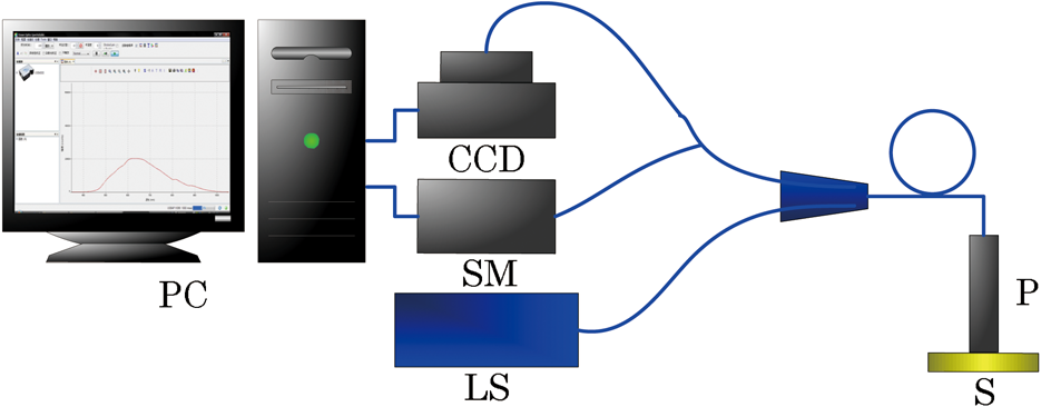 A total of 200 skin sites from 10 volunteers, which included six Chinese, three Indians, and one Caucasian, were measured in this experiment. A leave-one-out cross validation strategy12 was used to analyze the data to evaluate the modified Wiener estimation method in an unbiased manner. The reason for using human skin measurements for calibration instead of color chart as in several other previous studies was that such calibration data similar to the test data should yield better accuracy in estimated results.13 The proposed method was coded and run in Matlab (R2008a, MathWorks, U.S.). A total of four methods, which include the traditional Wiener estimation and the modified Wiener estimation methods using three different strategies for correcting estimated RGB values, were utilized to estimate diffuse reflectance spectra from given RGB values, and their performance was compared against each other. The mean root mean square errors (RMSE) of estimated diffuse reflectance spectra relative to measured diffuse reflectance spectra for four methods are shown in Table 1. The representative spectra for the modified Wiener estimation method with the best and worst accuracy are shown in Fig. 4. Table 1Comparison of mean RMSE between the traditional Wiener estimation (WE) and modified WE.
Fig. 4Comparison between the measured diffuse reflectance spectrum and the spectrum estimated by the modified Wiener estimation method in the cases of (a) best estimation and (b) worst estimation. The results from traditional Wiener estimation are also shown for comparison.  Table 1 shows that the modified Wiener estimation method reduces the RMSE by 7.2% or 4.5%, respectively, compared to the traditional Wiener estimation, when strategy 1 or 2 is used to correct new RGB values. Moreover, a much larger reduction in the RMSE, i.e., 15.3%, is seen when the selection step is added. This implies that more accurate new color values yielded by the selection step are critical to the accurate estimation of diffuse reflectance spectra. It took 19.3 milliseconds to estimate the diffuse reflectance spectra from 400 to 700 nm with a resolution of 5 nm at 50 skin sites in a PC with Intel Core 2 CPU 2.4 GHz, 2 G RAM and Windows Vista operating system. This short estimation time implies the potential of performing a real-time estimation of diffuse reflectance spectra based on color images acquired from a large tissue area. In conclusion, we have developed a modified Wiener estimation method by synthesizing new colors from the system matrix based on a calibration data set. The proposed method significantly improved the accuracy of estimated diffuse reflectance spectra for RGB color values acquired from human skin. This method is fast; thus, it may allow for real-time estimation of diffuse reflectance spectra from a large tissue area when combined with color imaging, which provides a cost-effective alternative to spectral imaging, with the additional advantage of high spectral resolution. AcknowledgmentsThe authors would like to acknowledge the financial support from a Tier 1 grant (Grant No. RG47/09) and a Tier 2 grant (Grant No. MOE2010-T2-1-049) funded by the Ministry of Education in Singapore. ReferencesQ. Liu,
“Role of optical spectroscopy using endogenous contrasts in clinical cancer diagnosis,”
World J. Clin. Oncol., 2
(1), 50
–63
(2011). http://dx.doi.org/10.5306/wjco.v2.i1.50 Google Scholar
A. A. StratonnikovV. B. Loschenov,
“Evaluation of blood oxygen saturation in vivo from diffuse reflectance spectra,”
J. Biomed. Opt., 6
(4), 457
–467
(2001). http://dx.doi.org/10.1117/1.1411979 JBOPFO 1083-3668 Google Scholar
J. SwartlingJ. S. DamS. Andersson-Engels,
“Comparison of spatially and temporally resolved diffuse-reflectance measurement systems for determination of biomedical optical properties,”
Appl. Opt., 42
(22), 4612
–4620
(2003). http://dx.doi.org/10.1364/AO.42.004612 APOPAI 0003-6935 Google Scholar
C. Zhuet al.,
“Diagnosis of breast cancer using fluorescence and diffuse reflectance spectroscopy: a Monte-Carlo-model-based approach,”
J. Biomed. Opt., 13
(3), 034015
(2008). https://doi.org/http://link.aip.org/link/doi/10.1117/1.2931078 JBOPFO 1083-3668 Google Scholar
S. Barontiet al.,
“Multispectral imaging system for the mapping of pigments in works of art by use of principal-component analysis,”
Appl. Opt., 37
(8), 1299
–1309
(1998). http://dx.doi.org/10.1364/AO.37.001299 APOPAI 0003-6935 Google Scholar
A. A. Wagadarikaret al.,
“Video rate spectral imaging using a coded aperture snapshot spectral imager,”
Optic. Express, 17
(8), 6368
–6388
(2009). http://dx.doi.org/10.1364/OE.17.006368 OPEXFF 1094-4087 Google Scholar
J. Y. Hardeberg,
“Acquisition and reproduction of color images: colorimetric and multispectral approaches,”
Ecole Nationale Superieure des Telecommunications,
(1999). Google Scholar
M. ShiG. Healey,
“Using reflectance models for color scanner calibration,”
J. Opt. Soc. Am. A, 19
(4), 645
–656
(2002). http://dx.doi.org/10.1364/JOSAA.19.000645 JOAOD6 0740-3232 Google Scholar
H. Haneishiet al.,
“System design for accurately estimating the spectral reflectance of art paintings,”
Appl. Opt., 39
(35), 6621
–6632
(2000). http://dx.doi.org/10.1364/AO.39.006621 APOPAI 0003-6935 Google Scholar
H. L. Shenet al.,
“Reflectance reconstruction for multispectral imaging by adaptive Wiener estimation,”
Optic. Express, 15
(23), 15545
–15554
(2007). http://dx.doi.org/10.1364/OE.15.015545 OPEXFF 1094-4087 Google Scholar
Q. LiuN. Ramanujam,
“Scaling method for fast Monte Carlo simulation of diffuse reflectance spectra from multilayered turbid media,”
J. Opt. Soc. Am. A, 24
(4), 1011
–1025
(2007). http://dx.doi.org/10.1364/JOSAA.24.001011 JOAOD6 0740-3232 Google Scholar
J. S. U. Hjorth,
“Cross validation,”
Computer Intensive Statistical Methods: Validation Model Selection and Bootstrap, 27
–28 Chapman & Hall, London
(1994). Google Scholar
H. L. ShenJ. H. Xin,
“Spectral characterization of a color scanner based on optimized adaptive estimation,”
J. Opt. Soc. Am. A, 23
(7), 1566
–1569
(2006). http://dx.doi.org/10.1364/JOSAA.23.001566 JOAOD6 0740-3232 Google Scholar
|


