
|
|
|
Medical imaging has played an important role in exploring physiology and diagnosing disease. Optical imaging has the advantages of use of nonionizing radiation, low cost, optical contrast sensitive to physiological parameters, and access to various optical contrast agents. Owing to strong light scattering, however, purely optical imaging suffers from either shallow penetration or low resolution. Photoacoustic (PA) imaging, based on the detection of laser-induced internal acoustic waves, was designed to take advantage of optical absorption contrast yet achieve ultrasonic resolution. This technology can provide an imaging depth up to several centimeters in tissue.1 PA imaging can be implemented in the mode of either computed tomography or direct image formation. The former mode has been developed and employed to image the brain cortex and whole head of small animals and has provided high spatial resolution and low imaging artifacts.2, 3 This mode, however, has relatively poor out-of-plane (elevation) resolution. The latter mode is based on a focused ultrasonic transducer. In this mode, focusing provides transverse (lateral) resolution, whereas temporal resolution furnishes axial (depth) resolution.4 Since bright-field illumination creates strong interference signals from tissue surfaces, Maslov developed dark-field reflection-mode photoacoustic microscopy (PAM) to minimize the interference.5 The initial implementation of PAM was based on a high-frequency ultrasonic transducer providing high spatial resolution. However, the imaging depth was limited to . In this letter, as well as our conference report,6 we present a scaled-up deep reflection-mode PA imaging system. The goal is to image much deeper than the limit while the depth pixel count, defined as the ratio of the imaging depth to the axial resolution, is maintained at more than 100. The capability of deeper imaging enables noninvasive mapping of deep structures such as the spleen of relatively heavy, small animals. The scaled-up reflection-mode PA imaging system is shown schematically in Fig. 1 . The dark-field ring-shaped illumination is formed by a concave lens, a spherical conical lens, and an optical condenser in tandem. This illumination has a great advantage over bright-field illumination in that it can reduce the generation of surface PA waves and improve the detection of deep PA waves. The prisms mounted on an XY-linear translation stage enable higher optical energy delivery than the optical fibers used in the high-frequency PAM system. To achieve deep penetration of light, we chose the near-infrared wavelength for the excitation. This wavelength is an isosbestic point of the molar extinction spectra of oxy- and deoxy-hemoglobin. The light source is a tunable Ti:sapphire laser (LT-2211A, LOTIS TII) pumped by a Q-switched Nd:YAG laser (LS-2137/2, LOTIS TII). The laser system provides light pulses of a pulse duration with a pulse repetition rate. The light beam is sufficiently broadened to conform to the maximum permissible exposure limit for the skin at this wavelength .7 To receive deep PA signals with minimal ultrasonic attenuation, we chose a central frequency for the ultrasonic transducer (V308, Panametrics-NDT). This transducer is spherically focused with a focal length, a 1.91-cm-diam active element, and a 72% nominal bandwidth. The use of a highly focused transducer (f-number: 1.33) is important for achieving good transverse resolution. The transducer is immersed in a water tank that has a opening sealed with a thin clear membrane. A subject is scanned through this opening while acoustically coupled with acoustic gel (Ultrasound Scanning Gel, Sonotech, Inc.). The PA signals are amplified by an amplifier (5072PR, Panametrics-NDT) and digitized and averaged by an oscilloscope (Tektronix TDS 5054). Signals from a photodiode (DET110, Thorlabs) are used to compensate for pulse-to-pulse fluctuations in laser energy. A computer controls the XY-linear translation stage for raster scanning and stores all the signals. We estimated the peak-to-peak signal to root-mean-square noise ratio (SNR), axial resolution, and transverse resolution as a function of the imaging depth by imaging human hair fibers [Figs. 2a, 2b, 2c ] in a 10% porcine gelatin containing 1% Lyposyn II. The reduced scattering coefficient of this phantom8 was , which is close to that of biological tissues. At the depth (the working distance of the transducer), images averaged 5 times achieved a SNR, a axial resolution, and a transverse resolution. The transverse resolution matches approximately the product of the f-number (1.33) and the central acoustic wavelength ( , obtained from the PA signals). Therefore, the pixel count in the depth direction was 136, greater than the targeted 100. Compared with the resolution of micro-CT (20 to ) and micro-MRI (100 to ), the axial resolution of the proposed system is similar. However, the imaging depth is less than those of both CT and MRI. PA imaging, however, is sensitive to intrinsic functional optical contrast. Figure 2d demonstrates the possible maximum imaging depth in chicken breast tissue. We imaged two -diam horse hairs located and deep, where the SNRs were measured to be and , respectively, with 30 times averaging. Synthetic-aperture focusing and coherence weighting were applied to improve the transverse resolution as well as the SNR.9 The rubber plate at a depth was also imaged. Fig. 2(a) SNR versus imaging depth (depth of the ultrasonic focal point). (b) Axial resolution versus imaging depth. (c) Transverse resolution versus imaging depth. (d) Demonstration of the maximum imaging depth in chicken breast tissue. 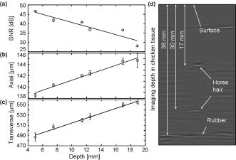 Internal organ imaging in both small and large animals is a valuable application of deep reflection-mode PA imaging. Internal organs such as the spleen have not been imaged reliably thus far using PA imaging techniques. Figure 3a shows the maximum amplitude projection (MAP) of the PA image of a spleen and a stomach of the rat weighing with the skin intact post mortem. Figure 3b shows the corresponding invasive anatomical photograph of the rat spleen obtained after PA imaging. The corresponding B-scan images in Fig. 3c show the depth information of the spleen and the stomach. The spleen was located to deep, and the adjacent stomach was even deeper. A three-dimensional (3-D) volumetric image is shown in Fig. 3d, which reveals the contour of the spleen and its depth. Fig. 3Noninvasive PA image of the spleen of a rat with the skin intact post mortem. (a) Noninvasive MAP image of the spleen and the stomach of the rat. (Color map was adjusted to show more details of the spleen by saturating the high intensity at the stomach.) (b) Corresponding invasive anatomical photograph. (c) B-scan image corresponding to the dashed line in (a) showing the depth information of the spleen and the stomach. (d) Three-dimensional (3-D) volumetric image showing the shape of the spleen and its depth.  In summary, a deep reflection-mode PA imaging system was successfully constructed using dark-field illumination and a highly focused ultrasonic transducer. The imaging depth of this system was proven to be up to in chicken breast tissue at the 804-nm wavelength. We imaged the spleen and the stomach of a rat, which have not been explored widely by PA imaging. In the future, continuous scanning and the use of a laser with a higher pulse repetition rate will be used to accelerate the data acquisition. AcknowledgmentsWe are grateful to Konstantin Maslov and Xinmai Yang for experimental assistance. This research is sponsored in part by National Institutes of Health Grant Nos. R01 EB000712 and R01 NS46214 (BRP). ReferencesG. Ku and
L. V. Wang,
“Deeply penetrating photoacoustic tomography in biological tissues enhanced with an optical contrast agent,”
Opt. Lett., 30 507
–509
(2005). https://doi.org/10.1364/OL.30.000507 0146-9592 Google Scholar
X. Wang,
Y. Pang,
G. Ku,
X. Xie,
G. Stoica, and
L.-H. V. Wang,
“Noninvasive laser-induced photoacoustic tomography for structural and functional in vivo imaging of the brain,”
Nat. Biotechnol., 21 803
–806
(2003). https://doi.org/10.1038/nbt839 1087-0156 Google Scholar
K. H. Song,
G. Stoica, and
L. V. Wang,
“In vivo three-dimensional photoacoustic tomography of a whole mouse head,”
Opt. Lett., 31 2453
–2455
(2006). 0146-9592 Google Scholar
R. G. M. Kolkman,
E. Honderbrink,
W. Steenbergen, and
F. F. M. de Mul,
“In vivo photoacoustic imaging of blood vessels using an extreme-narrow aperture sensor,”
IEEE J. Sel. Top. Quantum Electron., 9 343
–346
(2003). https://doi.org/10.1109/JSTQE.2003.813302 1077-260X Google Scholar
K. Maslov,
G. Stoica, and
L.-H. V. Wang,
“In vivo dark-field reflection-mode photoacoustic microscopy,”
Opt. Lett., 30 625
–627
(2005). https://doi.org/10.1364/OL.30.000625 0146-9592 Google Scholar
K. H. Song and
L. V. Wang,
“Deep reflection-mode photoacoustic imaging and resolution scalability with depth,”
Proc. SPIE, 643773
(2007). 0277-786X Google Scholar
, “American National Standard for the safe use of lasers,”
(2000) Google Scholar
S. T. Flock,
S. L. Jacques,
B. C. Wilson,
W. M. Star, and
M. J. C. van Gemert,
“Optical properties of Intralipid: A phantom medium for light propagation studies,”
Lasers Surg. Med., 12 510
–519
(1992). https://doi.org/10.1002/lsm.1900120510 0196-8092 Google Scholar
M.-L. Li,
H. Zhang,
K. Maslov,
G. Stoica, and
L.-H. Wang,
“Improved in vivo photoacoustic microscopy based on a virtual detector concept,”
Opt. Lett., 31 474
–476
(2006). https://doi.org/10.1364/OL.31.000474 0146-9592 Google Scholar
|


