
|
|
1.IntroductionIndocyanine green (ICG) is a tricarbocyanine dye that absorbs and fluoresces in the near-IR (NIR) wavelength region1, 2 (650 to ). It is used extensively in clinical settings to measure cardiac output and plasma volume, image choroidal structure, and assess liver function.3, 4 The minimal toxicity and vascular retention of ICG have motivated investigations into its utility for diagnostic purposes and a variety of therapeutic applications including photodynamic therapy, tissue welding, and photothermal therapy. 5, 6, 7, 8, 9, 10, 11, 12, 13, 14 As water and most intrinsic biomolecules within tissue do not absorb strongly within the NIR range,15 optical targeting of exogenously administered ICG may enable the selective treatment of deeper tissue structures. Our previous studies have demonstrated that ICG is a useful exogenous chromophore for laser-mediated destruction of blood vessels within rabbit earlobes as a model system for hypervascular lesions.16 After a bolus injection, the concentration of ICG within the bloodstream drops at an exponential rate, with a half time ranging between 2 and . ICG binds readily to plasma proteins and is rapidly excreted to bile.1, 17 The inability to localize freely dissolved ICG to a desired target and its relatively short retention time within the target limits its potential for diagnostic and therapeutic applications. Moreover, ICG effectiveness is hindered by its molecular instability, which causes18, 19 its optical properties to vary with factors such as concentration, temperature, solvent medium, and pH. The absorbance spectrum of ICG varies considerably with concentration. ICG molecules in solution remain in monomeric form at concentrations below approximately , and the absorbance spectrum shows a peak between 780 and , depending on the solvent medium.18 At higher concentrations, ICG forms dimers and oligomers, resulting in a new, lower absorbance peak between 680 and , again dependent on the solvent medium. At even higher concentrations (greater than ), ICG is prone to J-aggregation, a process in which the molecules self-organize over time into a highly aggregated, thread-like arrangement. The presence of J-aggregates results20, 21 in increased absorbance at a new peak of . ICG is known to undergo a variety of physicochemical transformations in aqueous media. It degrades over time through the formation of leucoforms, a molecular species with less conjugation, resulting in diminished absorbance in the IR range, discoloration, and a shift in peak absorbance.18, 20, 21 The degradation process occurs more readily in response to light exposure or elevated temperatures.22 In an effort to overcome the drawbacks faced with freely dissolved ICG, researchers are currently investigating techniques to encapsulate ICG into nanometer-sized carrier particles.23, 24, 25 Encapsulation within these nanoparticles has the potential to modulate the circulation kinetics of drugs, and by functionalizing the particle surface, therapeutic agents could be localized to targeted locations in the body. Researchers have reported on the use of ICG encapsulated in oil-water emulsion particles as a chromophore for photothermal heating.23, 24 Other investigators have encapsulated ICG at lower concentrations in biocompatible poly ( , -lactide-co-glycolide) (PLGA) nanoparticles for fluorescence contrast imaging.25 We have previously reported26, 27 on the synthesis and characterization of nanoparticle-assembled capsules (NACs) containing ICG. With particle diameters less than , the NACs contain a polymer-matrix core consisting of positively charged polymer molecules, poly-(allylamine hydrochloride) (PAH), bridged by negatively charged multivalent phosphate salt molecules. A coating of silica nanoparticles surrounded the core. Through charge interactions, ICG molecules are embedded within the polymer-salt matrix core and through the capsule surface. When compared to other encapsulation methods, the NACs offer the advantage of easy synthesis process, size controllability, and encapsulation efficiency. Incorporating ICG into the NAC delivery capsules gives rise to a potentially localizable photothermal absorbing system suitable for laser heating of abnormal tissue structures such as superficial tumors. In this study, we incorporate ICG into a modified version of the NAC capsule system in which the silica nanoparticle coating is replaced by dextran polymer, allowing for increased biocompatibility and functionalization.28 Our capsule diameters range between 400 and , smaller than the diameter range of microcapsules and yet larger than the range of particles conventionally referred to as nanoparticles . Consequently, we employ the term “mesocapsule” (MC) to refer to these dextran capsules to reflect their intermediate diameter range. The dextran outer coating and polymer-salt matrix core in our MC system limit the ability of the ICG molecules to interact with plasma proteins, and therefore could potentially slow down the excretion kinetics from the vasculature. We envision that the MC system can be used to accumulate ICG at the targeted location. On subsequent exposure to high pH or temperature, the MCs can be disassembled, liberating ICG within the microenvironment of the target. We focus on determining how encapsulating ICG within MCs can influence the sensitivity of ICG to temperature and light exposure. Specifically, absorbance measurements were recorded on (1) ICG released from disassembled MCs and (2) freely dissolved ICG to assess how encapsulation affected the absorbance spectrum. The degradation kinetics of both systems at different temperatures and when exposed to intense broadband light and different temperatures were evaluated. 2.Materials and Methods2.1.MC PreparationICG was purchased in powder form under the product name Cardiogreen from Sigma. Stock solutions of were prepared by dissolving the powder form into sterile, triple-distilled water, covered with foil, and refrigerated at to minimize degradation. ICG-containing MCs were prepared using a simple three-step process. The initial step involved the development of a spherical polymer-salt matrix by lightly mixing poly- (allyamine hydrochloride) (PAH, in deionized , ) with disodium phosphate ( , , , ) for in a centrifuge tube. Through ionic crosslinking, a cationic polymer-salt aggregate was formed between the positively charged PAH polymers and the anions. The ratio of total negative charge of the added salt to the total positive charge of the polymer, denoted as the ratio, was 6, satisfying the necessary conditions for aggregate formation.26, 27 The second step followed immediately after aggregate formation. The aggregate suspension was mixed with aqueous ICG solution ( , ) for 30 s. By electrostatic attraction and hydrophobic interaction, the negatively charged ICG molecules penetrated into the aggregates. By adding a more dilute solution of ICG ( , ) in, a batch of MCs containing less ICG was also prepared for the light exposure studies. These MCs with less ICG were compared with a diluted preparation of ICG solution. In the final step, an additional 0.5 ml of dextran polymer ( , purchased from Aldrich, in ) was added to the mixture and mixed for . The dextran molecules formed a coating around the surface of the aggregate through electrostatic interaction and H-bonding. The capsules were allowed to age for , at which point they were centrifuged at for and resuspended in phosphate-buffered saline (PBS) by sonication. The process was repeated twice to remove excess precursors. The resultant MCs remained suspended in PBS, with pH close to physiological levels in blood , for the duration of the experiments. For comparison with the MCs, ICG aqueous solution was made by diluting the ICG stock solution with sterile PBS. A concentration of was chosen to match the ICG concentration contained in the MC suspensions. More dilute ICG solution was made to match the ICG concentration in the less concentrated MC preparations by further diluting down to . 2.2.Absorbance SpectroscopyAbsorbance measurements were taken from 400 to using a spectrophotometer (Shimadzu UV-2401PC) equipped with a tungsten excitation lamp. For each absorbance reading, of sample was extracted and placed in an Eppendorf tube. Immediately before taking the absorbance measurement, of was added to each sample to increase the pH. Increasing the pH served two purposes. Earlier experiments revealed that, at a higher pH , the MCs lost their integrity and ICG was liberated back into solution. Disassembling the capsules eliminated the effect of Mie scattering by the submicrometer-diameter MCs. Additionally, it was found that the nonlinear relationship between ICG concentration and its absorbance spectrum, which results from ICG’s tendency to form molecular aggregates at higher concentrations, was eliminated over a wide concentration range at higher pH (unpublished results). After adding , of the sample was removed and further diluted in of PBS in a disposable cuvette. PBS was used as a reference for all absorbance measurements. Stability of ICG at high pH was also assessed. Preparations of ICG in PBS at high pH and concentrations ranging from 5 to were stored in the dark at room temperature. Absorbance profiles of these samples were recorded at , 20, 40, and . No differences in the absorbance profiles were observed up to after preparation. By taking a standard curve, the relationship between peak absorbance value and ICG concentration was found to be linear over the range of concentrations used in this study, from 1.25 to . 2.3.Calculation of Encapsulation EfficiencyTo measure the encapsulation efficiency of ICG within the MCs, a standard absorbance profile was created from the peak absorbance values of several samples of freshly dissolved ICG in PBS at different concentrations. The mass of ICG encapsulated within the MCs was determined by measuring the absorbance value of supernatants and disintegrated MCs and relating the measurements to their corresponding ICG concentrations. It was assumed that the ICG monomers did not degrade during the ICG-MC synthesis. We determined as 2.4.Size Analysis and Morphology CharacterizationParticle size analysis was conducted using brightfield and fluorescence microscopy. Small volumes of MCs suspended in PBS were pipetted onto a glass slide and covered with a coverslip. Scanning electron microscopy was used to characterize the MC morphology. 2.5.Light SensitivityEffect of light exposure on ICG absorbance characteristics was analyzed for MCs containing ICG and freely dissolved ICG at room temperature. Preparations were pipetted into Petri dishes ( per trial) placed directly beneath a broadband tungsten lamp (Lowel Prolite). Optical power was measured using a Coherent Lasermate 10 power meter with thermal power head. Using an adjustable iris and a collimating lens, the entire dish was illuminated at a uniform intensity of . With this intensity and exposure duration, the total energy delivered is comparable to light doses used for photothermal therapy.29 At various time points for up to , was withdrawn from the Petri dish and the absorbance spectrum was recorded. The temperature of the sample was monitored using a thermometer and never exceeded . Each experiment was conducted in triplicate. 2.6.Temperature SensitivityPreparations of freely dissolved ICG and MCs were aliquotted into microcentrifuge tubes ( per tube) and remained covered in foil. The samples were either refrigerated at , maintained at room temperature , or incubated in a water bath at . Every , the absorbance spectra for the preparations were recorded and analyzed for up to . The different temperatures were used to investigate the influence of temperature on the optical stability of ICG. Specifically, incubation at for several days was used to simulate physiological conditions experienced by nanoparticles as they accumulate at a targeted tumor site following a bolus injection. To determine whether ICG escaped from the MCs over the course of our experiments, the capsules were centrifuged and resuspended in PBS. The absorbance profiles were recorded for both the MCs and the supernatant. Three samples were used for each time point. 2.7.Absorbance CalculationsAlterations in absorbance spectra are a measure of the changes in the concentration of nondegraded ICG molecules as a function of time and wavelength . We use equations reported by Holzer to quantitatively express: where and represent the absorbance values immediately after synthesis and after elapsed time , respectively, represents the initial concentration of nondegraded ICG molecules, represents the concentration of degraded molecules, and are the, respective, molar absorptivity of nondegraded and degraded ICG, and (in centimeters) represents the path length of the sample.18 From these relationships, the parameter is derived to represent the percent change between the initial and final absorbance spectrum:The ratio provides a means to evaluate which molecular species, monomers, dimers, or other aggregates within the ICG solutions are most sensitive to light exposure. It is assumed that the degraded molecules have minimal absorption near the monomeric and dimeric peaks of nondegraded ICG molecules18, 20 [ , and , respectively].ICG is useful for various diagnostic and therapeutic applications in its native, monomeric state. Therefore, we are interested in characterizing the potential spectral changes in the absorption peak of ICG monomers at that may result from external physical stimuli such as temperature and intense, broadband light exposure, and define the parameter as 2.8.Degradation KineticsThe change in absorbance over time was used to determine the kinetics of ICG degradation. As the peak absorbance value was linearly proportional to the ICG concentration within the concentration range used in this study, the decrease in absorbance peak over time was assumed to reflect the physicochemical transformations of ICG molecules. The degradation processes were assumed to occur in a single step, and degree of linear fits was used to determine the relationship between the degradation rate and the concentration of ICG. Linear fits for versus time, versus time, and for versus time, were calculated to determine whether the rate of ICG monomer degradation was independent of ICG concentration (zero order), directly proportional to ICG concentration (first order), or proportional to the square of ICG concentration (second order). The reaction order that yielded the best correlation coefficient was considered to be the approximate order. 3.Results3.1.MC MorphologyFigure 1 shows a scanning electron microscopy (SEM) image of the dextran-coated MCs. The MCs are roughly spherical, and ranged from 400 to in diameter. Brightfield and fluorescence microscopy verified that the MCs did not aggregate appreciably in aqueous solution. 3.2.Encapsulation Efficiency and ICG Release KineticsFreshly prepared MCs were disintegrated with , and was evaluated by comparing the peak absorbance values to a standard curve for ICG dissolved in PBS. Our earlier experiments revealed that depends largely on the initial ICG concentration and volume utilized in the MC synthesis process. The ICG concentration and volume used for these studies was selected to optimize , which was determined to range between 48 and 85%. Other experiments showed that when not encapsulated as MCs, PAH, phosphate, and dextran in solution had negligible influence on the absorbance profile of ICG. The amount of ICG released from the MCs over the course of our studies was determined by absorbance measurements of the supernatants from preparations stored in dark at 3, 22, and over 2 or 4 days. In the case of MCs stored at 3 and at , the ICG concentration within the supernatants did not exceed 3% of the initial ICG concentration within the capsules. At , the supernatant concentration did not exceed 7.5% of the initial capsule concentration. These results suggest that more than 90% of ICG did not escape from the MCs over the course of our experiments. ICG leakage was not considered in the light sensitivity and temperature sensitivity studies. The ICG molecules remain trapped within the aggregate core by electrostatic interaction, and moreover, any change in the absorbance profile at later time points were attributable to the instability of the ICG molecules. 3.3.Light SensitivityFigure 2 shows the normalized change in monomeric peak absorbance value, , for both MCs and freely dissolved ICG in the dark when irradiated with broadband light for various exposure durations. At a concentration of , freely dissolved ICG was found to degrade significantly in response to broadband light exposure over the course of . MCs containing a comparable amount of ICG were found to degrade by a similar amount. Fig. 2Influence of broadband light exposure on, , normalized monomeric peak absorbance of freely dissolved ICG (left) and MCs (right) under different exposure conditions. Freely dissolved ICG and ICG within MCs experienced similar amounts of monomeric degradation at . At a more dilute concentration , ICG monomers within MCs were more stable, while monomers in freely dissolved ICG degraded much more significantly.  As the profiles of versus yielded the greatest correlation coefficients, the rate of degradation for peak absorbance, and therefore the degradation rate for nondegraded monomeric ICG, followed first-order kinetics. The rate constants were determined by the slope of the linear fits and are provided in Table 1 . Interestingly, the degradation rate constants are dramatically different for more dilute preparations (20% of the original concentration) that were irradiated with the same light exposure conditions. When compared to the typical ICG concentrations utilized for this study, the values in Table 1 reveal that the peak absorption value decreases much further for a dilute solution of freely dissolved ICG, with a degradation rate constant nearly 3 times higher than that of the typical solution. This is consistent with previous reports that demonstrate ICG’s increased sensitivity to light exposure at lower concentrations.18 Conversely, a more dilute suspension of MCs demonstrates greater photostability than the more concentrated preparations of MCs and freely dissolved ICG, evident by a more gradual slope. The results suggest that the protection of ICG from photodegradation provided by encapsulation within MCs is concentration dependent, with more dilute capsule preparations allowing greater stability. Figure 3 shows the profiles for samples that were exposed to broadband light for 120 minutes, the maximum exposure duration. We present results for the wavelength region between 600 and , as the absorbance outside this region was essentially negligible both before and after light exposure. In all cases, near maximal change in is found at the monomeric peak, , indicative of the monomers’ sensitivity to light-induced degradation. In the case of freely dissolved ICG at , the profile exhibits negative values at wavelengths greater than , suggesting that light exposure for resulted in increased molar extinction coefficient in this wavelength region. Other investigators have also reported18 increased absorbance at wavelengths greater than in response to laser light exposure for nearly . As it is assumed that the degraded ICG molecules have minimal absorbance at the monomeric and dimeric peak for nondegraded ICG, the values of and can be regarded as the fraction of ICG monomers and dimers that degraded over the course of the experiment, respectively. Approximately 83% of the monomers of the freely dissolved ICG in the dilute solution degraded, while roughly 50% of the monomers of the freely dissolved ICG and MCs degraded concentrations. In the case of the dilute suspension of MCs, 30% of the monomers degraded when exposed to light for . The freely dissolved ICG samples exhibited dramatic change at the monomeric peak in comparison to the dimeric peak. The flatter profiles of the ICG-MCs suggest that ICG monomers, aggregates, and other oligomer forms of the molecule exhibit comparable sensitivity to light induced degradation. Fig. 3Spectral absorbance ratio at of broadband light exposure for freely dissolved ICG (left) and MCs. Freely dissolved ICG degraded more readily at the more dilute concentration while the converse was observed in MCs.  The values at ( , ) for freely dissolved ICG at concentration of shows that approximately 20% of the dimers degraded, whereas nearly 70% of dimers degraded in the case of freely dissolved ICG at concentration of . The degradation of the monomeric and dimeric molecules resulted in discoloration of freely dissolved ICG at both concentrations. This discoloration was not as pronounced within the MC suspension. For the dilute MC suspension, only 26% of the dimers degraded, whereas 54% degraded in the MC suspension samples. 3.4.Temperature SensitivityTo evaluate how encapsulation of ICG within MCs affects ICG sensitivity to temperature, preparations of MCs and freely dissolved ICG at concentration of were incubated in the dark at 3, 22, and for up to . Figure 4 displays at various times for both systems at the three temperatures. In all cases, the correlation coefficients were , suggesting that the degradation kinetics did not suitably follow zero-, first-, or second-order trends. Nevertheless, the best-fit profiles for first-order kinetics are included in the figures. Freely dissolved ICG is far more vulnerable to thermal degradation than the ICG within the MCs, as the monomeric peak absorbance decreased by less than 10% for the MCs stored at after . Freely dissolved ICG also experienced noticeable degradation at 22 and , whereas virtually no degradation occurred for MCs stored at these temperatures over the course of the experiments. Figure 5 shows the normalized initial and final absorbance spectra of both freely dissolved ICG and ICG-MCs at incubated at for . The spectra are normalized by the initial monomeric peak value . The spectrum for freely dissolved ICG changed dramatically, exhibiting substantially reduced absorbance and broadening at the peak values as well as the emergence of a new, small peak near . However, the overall spectrum for the MCs retained the same shape, while the peak values diminished slightly. Similar, but less pronounced, changes in the monomeric and dimeric peaks were observed for the absorbance spectra of freely dissolved ICG incubated at 22 and , while the spectra for the ICG encapsulated in MCs had little change. Fig. 5Absorbance spectra normalized to the monomeric peak at for both freely dissolved ICG and MCs at of incubation at . Baseline profiles prior to incubation at are also presented for comparison. 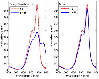 Figure 6 shows at three different temperatures. All of the spectra display large negative values in the NIR region . This might be attributable to the increase in absorbance over time that results from the first stages of J-aggregation. Reports have shown that, at room temperatures, J-aggregation of ICG molecules in solution does not set in for several weeks.20 However, because the initial absorbance in this region is so low , even a slight change is emphasized, i.e., normalizing by yields a more pronounced value for . With increasing temperature, ICG solution is more prone to degradation, as indicated by the higher values for . The maximal change for freely dissolved ICG occurs at the monomeric peak , indicating that the monomers degraded by 13, 25, and 42% when stored at 3, 22, and , respectively. ICG dimers also experience noticeable degradation, by as much as 12, 19, and 20% after being stored at 3, 22, and , respectively. The high values near indicate the degradation of dimers, although the actual local maximum occurs closer to . Fig. 6Spectral absorbance ratios for freely dissolved ICG and MCs, , at of incubation at different temperatures.  Table 1Calculated degradation rate constants for freely dissolved ICG and MCs in response to broadband light exposure; all constants have units of inverse minutes.
For the MCs, the maximal change in absorbance occurs around the dimeric peak in the samples stored at 3 and . The negative values of and suggest that more dimers are present after incubation at 22 and , respectively. At these temperatures, a slight negative change is also observed at the monomeric peak, however, the change is negligible and most likely a consequence of experimental error. For the sample stored at , the wavelengths at which there is the most pronounced change in absorbance occurs between 620 and , with a steady decline in sensitivity at higher wavelengths. At all three temperatures, is significantly lower for the MCs than for freely dissolved ICG stored under the same conditions. At all temperatures, less than 7% of the ICG monomers located within the MCs underwent degradation. 4.DiscussionDextran-coated electrostatically assembled MCs with diameters ranging from 400 to and containing ICG were synthesized with ranging between 48 and 85%. Synthesis parameters were selected to yield MCs with maximal values and smallest capsule diameter. Both and MC diameter depend largely on the initial ICG concentration and volume utilized in the synthesis process. Adding smaller amounts of concentrated ICG solution resulted in clumping of the polymer cores and therefore influenced the size of the MCs, while adding large volumes of more dilute ICG solution influenced the ratio of positive polymer to dissociated salt anions and resulted in smaller cores. The polymer aggregate core of the complex has a finite number of positive charges for ICG to bind to, which establishes a maximal limit for the number of ICG molecules that can be incorporated into each capsule. Encapsulating ICG molecules within MCs enables protection of the molecules from light-induced degradation only under certain conditions. The photostability of the complexes is dependent on their concentration. ICG monomers within the MCs remain resistant to degradation when the MCs are suspended in a larger volume of liquid (i.e., less dilute suspension of MCs). This is in contrast to freely dissolved ICG, whose stability decreases drastically in more dilute solutions. Our results show that entrapment within the MCs effectively protects ICG monomers from thermal degradation. The exposure to high temperatures causes a significant reduction in the absorbance of freely dissolved ICG at its monomeric and dimeric peaks, indicative of their degradation. At each temperature tested, the absorbance profile for the capsules at the final time shows a slight increase in absorbance at wavelength range from to , as seen for in Fig. 5. This could possibly indicate the temperature-independent onset of a molecular transformation such as J-aggregation within the MCs. Figure 6 also shows that a third constituent is prone to thermally induced changes for freely dissolved ICG stored at room temperature or warmer conditions. The molecule, which absorbs light between to , is most likely a more aggregated oligomer or possibly an unstable leucoform. The molecule is either not present in the ICG solution stored at , or it does not undergo any changes at this colder temperature. These phenomena must be further investigated. In Figs. 3 and 5, the large decrease of in the IR region for ICG solution reflect an increase in the absorbance value within this wavelength region. This could have emerged through charge transfer with ambient molecules, or it could indicate the formation of other photoproducts or aggregates that absorb light in the long-wavelength range. Photodegradation comes about from the electronic excitation of ICG molecules to a higher singlet or triplet energy state. The change in energy states can lead to a change in conformational state of the molecules, vibrational excitation, or slight heat generation from nonradiative relaxation. Any of these effects can lead to the formation of destructive radical species. Thermally mediated molecular degradation is attributable to the increased likelihood of vibrational and translational agitation, which also increases the prospect of radical formation or the diffusion of intrinsic solvent radicals within the solvent to vulnerable molecular sites.30 From our thermal stability results, it can be reasoned that, when encapsulated within the complexes, the mobility of ICG is significantly hindered by its charge interactions within the polymer-salt core. The electrostatic attraction immobilizes the molecules sufficiently enough to render them invulnerable to random changes in vibrational and translational states that occur at elevated temperatures. However, when the ICG molecules absorb photons of light, the excited molecule has sufficient energy to overcome the immobilizing electrostatic effect, and altered vibrational state and subsequent radical formation within the molecules can ensue. While, the effect of concentration on photostability must be further investigated, we presume that the effect could be explained by the density of ICG molecules and the amount of destructive radicals generated in response to light. In a more concentrated collection of capsules, the excited ICG molecules are more densely packed, and radicals generated from photo-excited molecules will have a more pronounced effect on nearby ICG molecules. A greater population of excited ICG molecules undergoing nonradiative relaxation also leads to a larger rise in temperature, which would more likely degrade the neighboring, vulnerable photoexcited ICG molecules. The same effect would not be found in more concentrated solutions of freely dissolved ICG, as they are more likely to produce aggregates and oligomers with different absorbance peaks. The discoloration and diminished absorbance of ICG at its NIR peak is attributable in part to fragmentation of molecules, but it is primarily the result of loss of conjugated double bonds within molecules that otherwise remain intact. Encapsulation within MCs reduces the likelihood of these molecular changes by immobilizing the molecules through electrostatic attraction to the positive charges distributed throughout the core. As the absorbance properties of ICG are not adversely affected through encapsulation within the MCs, the dextran-coated MCs containing ICG could be useful for photothermal therapy. The MCs can be used to overcome drawbacks that limit the ICG therapeutic abilities, including rapid clearance from the bloodstream and its inability to be localized. By functionalizing the dextran surface with site-specific antibodies, the MCs could provide an efficient means to deliver and localize ICG to targeted therapeutic sites such as tumors. 5.ConclusionICG was encapsulated within spherical dextran-coated MCs with diameters ranging from 400 to , and the sensitivity of the encapsulated ICG to broadband light and temperature was investigated. We found that the sensitivity of encapsulated ICG to light varies with the concentration of ICG incorporated within the MCs. Encapsulation of ICG within MCs reduces the effects of thermally induced degradation. AcknowledgmentsThis work was supported by a grant from the National Institutes of Health under Grant No. GMO 8362. We thank Dr. Parmeswaran Diagaradjane for several helpful discussions. ReferencesM. L. J. Landsman,
G. Kwant,
G. A. Mook, and
W. G. Zijlstra,
“Light-absorbing properties, stability, and spectral stabilization of indocyanine green,”
J. Appl. Physiol., 40 575
–583
(1976). 0021-8987 Google Scholar
S. A. Prahl,
“Optical absorption properties of indocyanine green (ICG),”
(2006) http://omlc.ogi.edu/spectra/icg/index.html Google Scholar
J. G. Webster,
“Measurement of flow and volume of blood,”
Medical Instrumentation: Application and Design, Wiley, New York (1998). Google Scholar
R. C. Benson and
H. A. Kues,
“Fluorescence properties of indocyanine green as related to angiography,”
Phys. Med. Biol., 23 159
–163
(1978). https://doi.org/10.1088/0031-9155/23/1/017 0031-9155 Google Scholar
V. Ntziachristos,
A. G. Yodh,
M. Schnall, and
B. Chance,
“Concurrent MRI and diffuse optical tomography of breast after indocyanine green enhancement,”
Proc. Natl. Acad. Sci. U.S.A., 97 2767
–2772
(2000). https://doi.org/10.1073/pnas.040570597 0027-8424 Google Scholar
A. Rübben,
S. Eren,
R. Krein,
H. Younoussi,
U. Böhler, and
V. Wienert,
“Infrared videoangiofluorography of the skin with indocyanine green—rat random cutaneous flap model and results in man,”
Microvasc. Res., 47 240
–251
(1994). https://doi.org/10.1006/mvre.1994.1018 0026-2862 Google Scholar
L. S. Bass,
N. Moazami,
J. Pocsidio,
M. C. Oz,
P. LoGerfo, and
M. R. Treat,
“Changes in type I collagen following laser welding,”
Lasers Surg. Med., 12 500
–505
(1992). https://doi.org/10.1002/lsm.1900120508 0196-8092 Google Scholar
S. Fickweiler,
R. M. Szeimies,
W. Bäumler,
P. Steinbach,
S. Karrer,
A. E. Goetz,
C. Abels,
F. Hofstädter, and
M. Landthaler,
“Indocyanine green: intracellular uptake and phototherapeutic effects in vitro,”
J. Photochem. Photobiol., B, 38 178
–183
(1997). https://doi.org/10.1016/S1011-1344(96)07453-2 1011-1344 Google Scholar
C. Abels,
S. Fickweiler,
P. Weiderer,
W. Bäumler,
F. Hofstädter,
M. Landthaler, and
R. M. Szeimies,
“Indocyanine green (ICG) and laser irradiation induce photooxidation,”
Arch. Dermatol. Res., 292 404
–411
(2000). 0340-3696 Google Scholar
W. R. Chen,
R. L. Adams,
A. K. Higgins,
K. E. Bartels, and
R. E. Nordquist,
“Photothermal effects on murine mammary tumors using indocyanine green and diode laser: an in-vivo efficacy study,”
Cancer Lett., 98 169
–173
(1995). 0304-3835 Google Scholar
W. Bäumler,
C. Abels,
S. Karrer,
T. Weiß,
H. Messmann,
M. Landthaler, and
R. M. Szeimies,
“Photooxidative killing of human colonic cancer cells using indocyanine green and infrared light,”
Br. J. Cancer, 80 360
–363
(1999). https://doi.org/10.1038/sj.bjc.6690363 0007-0920 Google Scholar
K. Urbanska,
B. Romnaowska-Dixon,
Z. Matuszak,
J. Oszajca,
P. Nowak-Sliwinska, and
G. Stochel,
“Indocyanine green as a prospective sensitizer for photodynamic therapy of melanomas,”
Acta Biochim. Pol., 49 387
–391
(2002). 0001-527X Google Scholar
C. Abels,
S. Karrer,
W. Bäumler,
A. E. Goetz,
M. Landthaler, and
R. M. Szeimies,
“Indocyanine green and laser light for the treatment of AIDS-associated cutaneous Kaposi’s sarcoma,”
Br. J. Cancer, 77 1021
–1024
(1998). 0007-0920 Google Scholar
V. V. Tuchin,
E. A. Genina,
A. N. Bashkatov,
G. V. Simonenko,
O. D. Odoevskaya, and
G. B. Altshuler,
“A pilot study of ICG laser therapy of acne vulgaris: photodynamic and photothermolysis treatment,”
Lasers Surg. Med., 33 296
–310
(2003). https://doi.org/10.1002/lsm.10211 0196-8092 Google Scholar
A. J. Welch,
M. J. C. V. Gemert,
W. M. Star, and
B. C. Wilson,
“Definitions and overview of tissue optics,”
Optical-Thermal Response of Laser Irradiated Tissue, 15
–46 Plenum, New York (1995). Google Scholar
M. A. Yaseen,
P. Diagaradjane,
B. M. Pikkula,
J. Yu,
M. S. Wong, and
B. Anvari,
“Photothermal and photochemical effects of laser light absorption by indocyanine green (ICG),”
Proc. SPIE, 5965 27
–35
(2005). 0277-786X Google Scholar
T. Desmettre,
J. M. Devoiselle, and
S. Mordon,
“Fluorescence properties and metabolic features of indocyanine green (ICG) as related to angiography,”
Surv. Ophthalmol., 45 15
–27
(2000). https://doi.org/10.1016/S0039-6257(00)00123-5 0039-6257 Google Scholar
W. Holzer,
M. Mauerer,
A. Penzkofer,
R. M. Szeimies,
C. Abels,
M. Landthaler, and
W. Bäumler,
“Photostability and thermal stability of indocyanine green,”
J. Photochem. Photobiol., B, 47 155
–164
(1998). https://doi.org/10.1016/S1011-1344(98)00216-4 1011-1344 Google Scholar
O. G. Bjornsson,
R. Murphy, and
V. S. Chadwick,
“Physicochemical studies of indocyanine green (ICG): absorbance/concentration relationship, pH tolerance and assay precision in various solvents,”
Experientia, 38 1441
–1442
(1982). https://doi.org/10.1007/BF01955757 0014-4754 Google Scholar
M. Mauerer,
A. Penzkofer, and
J. Zweck,
“Dimerization, J-aggregation and J-disaggregation dynamics of indocyanine green in heavy water,”
J. Photochem. Photobiol., B, 47 68
–73
(1998). https://doi.org/10.1016/S1011-1344(98)00205-X 1011-1344 Google Scholar
R. Philip,
A. Penzkofer,
W. Bäumler,
R. M. Szeimies, and
C. Abels,
“Absorption and fluorescence spectroscopic investigation of indocyanine green,”
J. Photochem. Photobiol., A, 96 137
–148
(1996). https://doi.org/10.1016/1010-6030(95)04292-X 1010-6030 Google Scholar
V. Saxena,
M. Sadoqi, and
J. Shao,
“Degradation kinetics of indocyanine green in aqueous solution,”
J. Pharm. Sci., 92 2090
–2097
(2003). https://doi.org/10.1002/jps.10470 0022-3549 Google Scholar
S. Mordon,
J. M. Devoisselle,
S. Begu, and
T. Desmettre,
“Laser-induced release of liposome-encapsulated dye: a new diagnostic tool,”
Lasers Med. Sci., 13 181
–188
(1998). 0268-8921 Google Scholar
T. J. Desmettre,
S. Soulie-Begu,
J. M. Devoisselle, and
S. R. Mordon,
“Diode laser-induced thermal damage evaluation on the retina with a liposome dye system,”
Lasers Surg. Med., 24 61
–68
(1999). https://doi.org/10.1002/(SICI)1096-9101(1999)24:1<61::AID-LSM10>3.3.CO;2-7 0196-8092 Google Scholar
V. Saxena,
M. Sadoqi, and
J. Shao,
“Enhanced photo-stability, thermal-stability and aqueous-stability of indocyanine green in polymeric nanoparticulate systems,”
J. Photochem. Photobiol., B, 74 29
–38
(2004). https://doi.org/10.1016/j.jphotobiol.2004.01.002 1011-1344 Google Scholar
J. Yu,
M. A. Yaseen,
B. Anvari, and
M. S. Wong,
“Synthesis of near-infrared absorbing nanoparticle-assembled complexes,”
Chem. Mater., 19 1277
–1284
(2007). https://doi.org/10.1021/cm062080x 0897-4756 Google Scholar
R. K. Rana,
V. S. Murthy,
J. Yu, and
M. S. Wong,
“Nanoparticle self-assembly of hierarchically ordered microcapsule structures,”
Adv. Mater. (Weinheim, Ger.), 17 1145
–1150
(2005). https://doi.org/10.1002/adma.200401612 0935-9648 Google Scholar
T. Heinze,
T. Liebert,
B. Heublein, and
S. Hornig,
“Functional polymers based on dextran,”
Adv. Polym. Sci., 205 199
–291
(2006). 0065-3195 Google Scholar
M. H. Niemz,
“Thermal interaction,”
Laser-Tissue Interactions: Fundamentals and Applications, 58
–85 Springer-Verlag, Berlin (1996). Google Scholar
J. F. Rabek, Photodegradation of Polymers: Physical Characteristics and Applications, Springer, Berlin (1996). Google Scholar
|
||||||||||||||||||||||||


