|
|
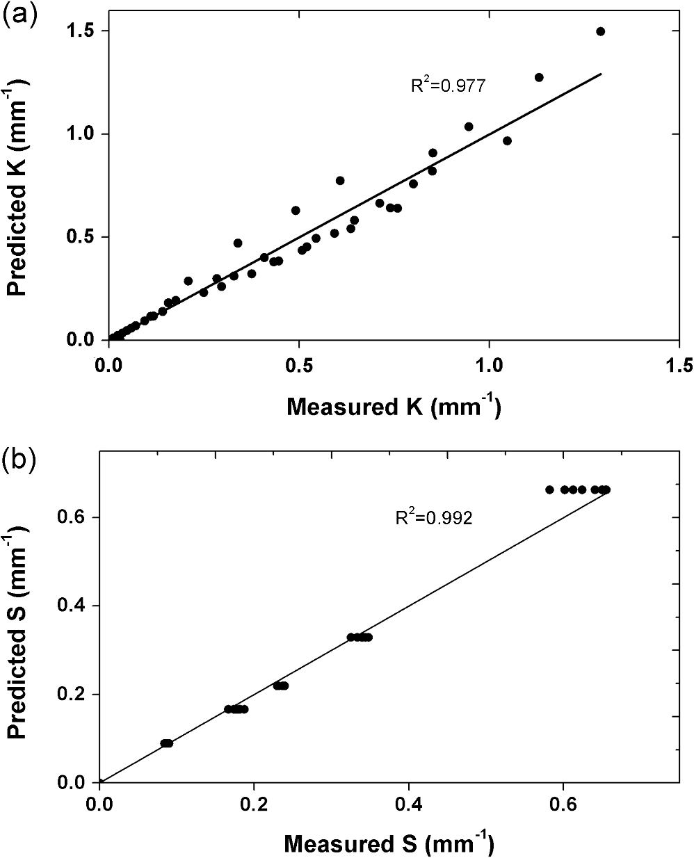
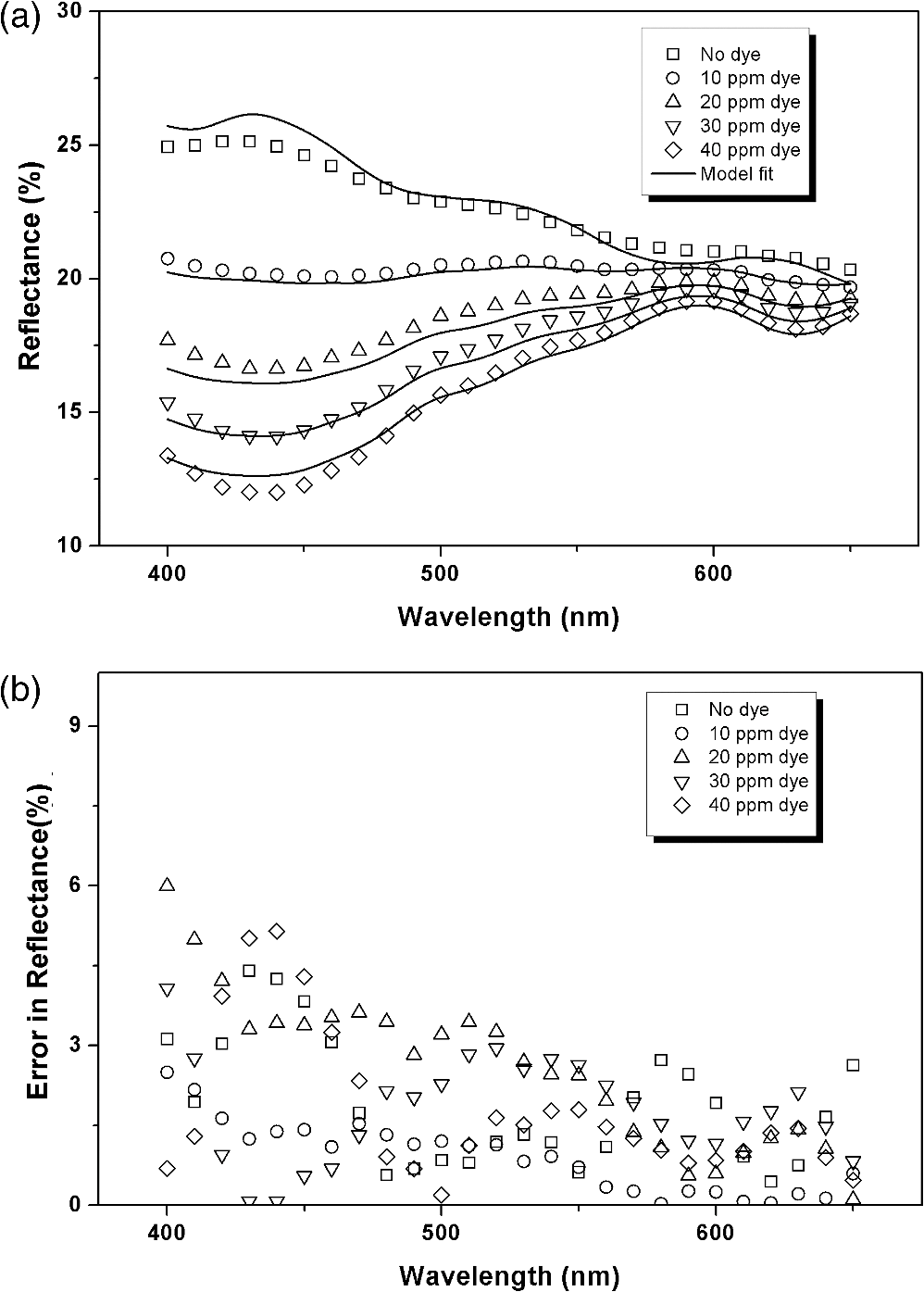
1.IntroductionA major challenge in tissue optics is to noninvasively characterize tissue properties for early diagnosis of disease. Optical properties such as scattering () and absorption () coefficients provide information regarding the morphological and biochemical composition of tissues. Various optical techniques are currently explored to obtain different parameters which represent tissue conditions. One such technique is diffuse reflectance spectroscopy,1–4 wherein the spectrum of light reflected from tissue is analyzed to obtain its absorption and scattering properties. Diffuse reflectance needs an accompanying numerical model to decipher the data for obtaining the optical parameters. This can be achieved by solving the radiative transfer equations5,6 in tissues. However, due to the complexity of the problem, no analytical solutions exist between the measured reflectance and the optical parameters. Accurate results can be obtained by using Monte Carlo methods,7,8 which are inherently very slow. Different authors9,10 have developed semi-empirical relations for extracting optical parameters from the reflectance spectra measured using fiber-optic probes with small source-detector separations. Diffusion approximation of the radiative transfer equation has been used by many authors to obtain optical parameters such as and reduced scattering coefficient () from the measured diffused reflectance spectrum.11,12 Kubelka–Munk (K-M) theory,13 one of the diffusion approximation-based theories, is widely used to obtain optical properties from the measured reflectance spectra. The advantage of this theory is that it provides simple analytical expressions for obtaining the optical parameters from the measured reflectance and transmittance. The reflectance and transmittance of a diffusive media of thickness is given by where , , and and are the K-M absorption and scattering coefficients, respectively.K-M coefficients are only phenomenological approximations, because the theory assumes incident radiation is diffuse and the scattering is isotropic. Similar to other diffusion approximation–based models, K-M theory also assumes higher scattering compared to absorption. Although these conditions are not completely met in many of the real systems, the theory nonetheless provides a simple quantitative way of describing light transport in diffused medium. Many authors14–17 have derived relations between the K-M coefficients and the more fundamental radiative transfer coefficients (, , and ). Here is the absorption coefficient, is the scattering coefficient, and is the anisotropy factor of the diffusive medium. In the diffusive approximation regime (), the general form of the relations between the K-M coefficients and the radiative transfer coefficients derived by different authors can be written as where is the reduced scattering coefficient. For an isotropic medium, Klier14 had shown that and , and the value of ranges from 0.5 to 1 and the value of ranges from to 3.33. van Gemert and Star15 extended this further to show that the K-M coefficients can be related to the reduced scattering coefficient in an anisotropic medium. For an anisotropic and highly scattering medium, they showed that and . However, in most of the measurement geometries, the condition that the incident beam is diffusive is not met. It is either fully collimated or partly collimated and partly diffusive. Various authors have extended this model further to include collimated incident flux and have shown that the K-M theory is also applicable in the case of collimated beam.18–20 Thennadil20 has shown that, for a collimated incident beam in the high scattering regime, depends only on scattering; however, the relation should be modified to include a function dependent on the anisotropy factor, .Here, we have developed an empirical relationship between the K-M and the radiative transfer coefficients for a collimated incident beam which is valid for both diffusive () and nondiffusive () regimes. This was achieved by measuring the total transmittance (collimated and diffuse) and total reflectance (diffuse and specular) of tissue phantoms with different set of optical parameters ( and ) and solving Eq. (1) to obtain the corresponding K-M coefficients and . The empirical relations between (, ) and (, ) were obtained, and their applicability for obtaining tissue optical parameters from measured reflectance spectra are discussed in this article. 2.Materials and Methods2.1.Tissue PhantomsLiquid tissue phantoms with different optical parameters were prepared in a 0.75-mm-thick glass cuvet by dispersing known concentrations of polystyrene microspheres and one of the two brown colored dyes in milliQ water. The Premium Dark Chocolate dye was procured from Devarsons Industries, India, and the Chocolate Brown TAS dye was procured from Roha Dyechem, India. Polystyrene microspheres of 0.5- and 1-μm diameter were procured from Duke Scientific, USA. Absorption coefficients of the dyes and the scattering coefficient () of the polystyrene spheres were measured using a Perkin Elmer Lambda900 UV-Vis spectrophotometer. The wavelength-dependent absorption coefficients of the two dyes for 10 ppm concentration are shown in Fig. 1. The anisotropy factor was calculated using a web-based Mie scattering calculator developed by Philip Laven.21 The reduced scattering coefficients were calculated using the relation . 2.2.Spectral MeasurementsTotal transmittance (collimated and diffuse) and total reflectance (specular and diffuse), at 488 nm, of the tissue phantoms were measured using an Ar-ion laser (Lexel, USA), a 90-mm-diameter integrating sphere (Labsphere, USA), and a spectrometer (ILT 900 from International Light, USA). The configurations used in these measurements are shown in Fig. 2(a) and 2(b). Reflectance was measured relative to a Spectralon® reflectance standard with a reflectance value of 99% in the 400- to 650-nm range. For reflectance measurement, the sample was held at 8 deg with respect to the direction of normal incidence, as shown in Fig. 2(a), to include the specular component of the reflected beam. The specular and the diffuse reflected light was collected by the integrating sphere and measured using the spectrometer. Reflectance from a water-filled reference cuvet was subtracted from the measured reflectance to obtain reflectance from the tissue phantom alone. During total transmittance measurement, the transmitted light was collected by an integrating sphere and measured using a spectrometer as shown in Fig. 2(b). A water-filled cuvet was used as reference during transmittance measurements to account for reflectance losses from the cuvet walls. Fig. 2Measurement configuration used to measure total reflectance (a) and total transmittance (b). (c) Measurement configuration of the commercial spectrophotometer used in validation measurements.  For the validation study, the wavelength-dependent reflectance spectra were measured using a handheld spectrophotometer (CM-2600d, Minolta, Japan). The configuration of the spectrophotometer is shown in Fig. 2(c). It is equipped with two pulsed Xenon lamps and a 52-mm-diameter integrating sphere. The sample was illuminated through an 8-mm-diameter aperture. The integrating sphere and the small aperture ensure that a broad collimated beam is incident on the sample. The reflected light is collected through the same aperture in the 8-deg viewing angle and is analyzed using a diffraction grating and a photodiode array. The reflectance was measured in the wavelength range of 400 to 650 nm with a spectral resolution of 10 nm. 3.Results3.1.Empirical Relations between K-M and Radiative Transfer CoefficientsThe method used to obtain the empirical relations between the radiative transfer and K-M coefficients is schematically shown in Fig. 3. First, the total reflectance () and total transmittance () values of a diffusive layer were measured for various sets of and . For a given set of and values, there is a unique set of and values that can be obtained by solving the coupled relations shown in Eq. (1). Finally, empirical relationships are obtained between the calculated sets of and values and the sets of radiative transfer coefficients ( and ) used in the tissue phantom. Fig. 3Schematic diagram of the method used to obtain empirical relations between radiative transport coefficients and the Kubelka–Munk (K-M) coefficients. From the measured total reflectance () and transmittance (), the K-M coefficients were calculated and related to the tissue phantom’s radiative transport coefficients ( and ). 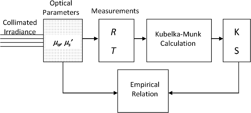 For and measurements, tissue phantoms with six different values—0, 0.22, 0.41, 0.54, 0.81, and —were obtained by varying the concentration of the 0.5-μm-diameter microspheres. For a constant , was varied from 0 to by varying the concentration of the Premium Dark Chocolate dye shown in Fig. 1. The measured and values for various values of and at 488 nm are shown in Fig. 4. For increasing , both and decrease. With increasing , decreases while increases. The measured and values are functions of and as shown in Eq. (1), which in turn depend on and . Fig. 4Measured values of total reflectance (a) and total transmittance (b) (at 488 nm) of tissue phantoms for various values of and . 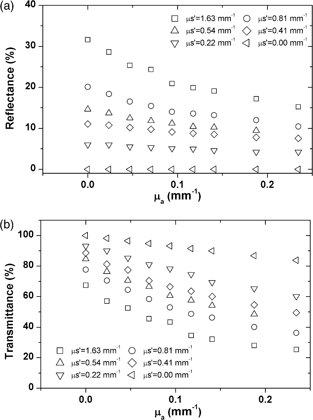 Earlier authors18–20 have shown that the K-M theory is also applicable to collimated incident beam in the regime where . In the current measurement geometry, the incident beam is collimated. However, not all the tissue phantoms fall in the regime . Assuming that K-M theory is applicable for all ranges of and , including , the values of and were calculated by solving the coupled equations in Eq. (1) for various values of and shown in Fig. 4. The calculated and values are plotted as a function of for constant values of and are shown in Fig. 5(a) and 5(b), respectively. Fig. 5K-M scattering coefficient (a) and K-M absorption coefficient (b) for tissue phantoms with various values of and . and were obtained from the measured total reflectance () and total transmittance values () by solving the K-M equations.  From Fig. 5(a), it is clear that depends strongly on and not on even in the diffusive regime . This is different from earlier reports which showed that the depends on and as shown in Eq. (2). Our measurement agrees with derivations of Gate17 and Thennadil20 where was zero. By fitting the experimental values to Eq. (2), we find the value of to be 0.408. Thus Figure 5(b) shows the dependence of as a function of and . Unlike , depends on both and . K-M absorption coefficient increases as increases. In the absence of scattering, is equivalent to the absorption coefficient . Increasing at a given further increases . In the presence of scattering, has an additional term which we can attribute to the absorption due to scattered photons. The absorption of the scattered photon depends not only on the absorption coefficient of the medium but also on the mean average distance traveled by the scattered photon, which is proportional to its scattering coefficient.10 A photon in a highly scattering medium will travel larger distances compared to a photon in a low-scattering medium. Hence the effective absorption of the scattered photon will depend on both the absorption coefficient and the scattering coefficient. We find that follows the following empirical relation: where and are expressed in units of . By fitting our data to the above empirical relation, we find the parameter values as and . Figure 6 shows predicted values of and using the empirical relations in Eqs. (3) and (4) against values of and calculated from measured and values using Eq. (1). We see an excellent fit, with values of 0.97, implying that our empirical relation is applicable to a wide range of and .3.2.Validation of Empirical Relations Using Tissue PhantomsTo validate the applicability of the empirical relations in Eqs. (3) and (4), tissue phantoms were prepared using 1-μm polystyrene spheres and five different concentrations (0, 10, 20, 30, and 40 ppm) of the Chocolate Brown TAS dye whose extinction coefficient is shown in Fig. 1. Total reflectance of the tissue phantoms was measured in the wavelength range of 400 to 650 nm using a commercial spectrophotometer as shown in Fig. 2(c). In this range, there is large variation of absorption as a function of wavelength. In certain regions ; in other regions . Because and of the tissue phantoms are known for all the wavelengths, and values can be calculated using Eqs. (3) and (4), and the reflectance for the entire wavelength range can then be predicted using Eq. (1). In Fig. 7(a), the measured reflectance spectra (dots) of tissue phantoms with various dye concentrations are shown along with the reflectance spectra predicted (solid lines) using Eqs. (3) and (4). Good agreement was obtained between the predicted and measured reflectance spectra. The errors in the predictions are shown in Fig. 7(b). The maximum error in prediction was about 6%, implying that the empirical relation between the optical parameters and K-M coefficient is valid over a wide range of optical parameters. 3.3.Extracting Optical Parameters from Measured ReflectanceBecause the empirical relations in Eqs. (3) and (4) are valid over wide range of optical parameters and wavelengths, it is possible to solve the inverse problem of extracting various optical parameters from the measured reflectance spectra. For example, parameters such as concentration of the chromophores and thickness of the scattering layer can be extracted simultaneously from the measured reflectance if the scattering parameters are known. This is feasible, since at different wavelengths, relations in Eqs. (3) and (4) are valid and independent of each other. One needs to solve for two unknown parameters using more than two equations. To verify the feasibility of simultaneously extracting dye concentration and layer thickness from the measured reflectance, tissue phantoms of two different thicknesses (450 and 750 μm) were prepared using 1000-nm polystyrene spheres and different concentrations of the Chocolate Brown TAS dye shown in Fig. 1. The reflectance of the tissue phantoms in the range of 400 to 650 nm was measured, and best fits to the reflectance curves were obtained using Eqs. (1), (3), and (4) by freely varying the two fitting parameters, namely the concentration of the dye and thickness of the phantom layer. The best fit obtained is shown in Fig. 8, and the parameters used to obtain the best fit are shown in Table 1 along with the actual parameters. The extracted concentration values were within an error of 2% to 10% whereas the extracted thickness values were within 2% error. Fig. 8Solving for optical parameters from the measured reflectance spectra of tissue phantoms. The best fits were obtained by varying two fitting parameters, namely dye concentration and layer thickness. The symbols show measured reflectance, and solid lines show the best fit from the model. The actual parameter values and the predicted values are shown in Table 1. 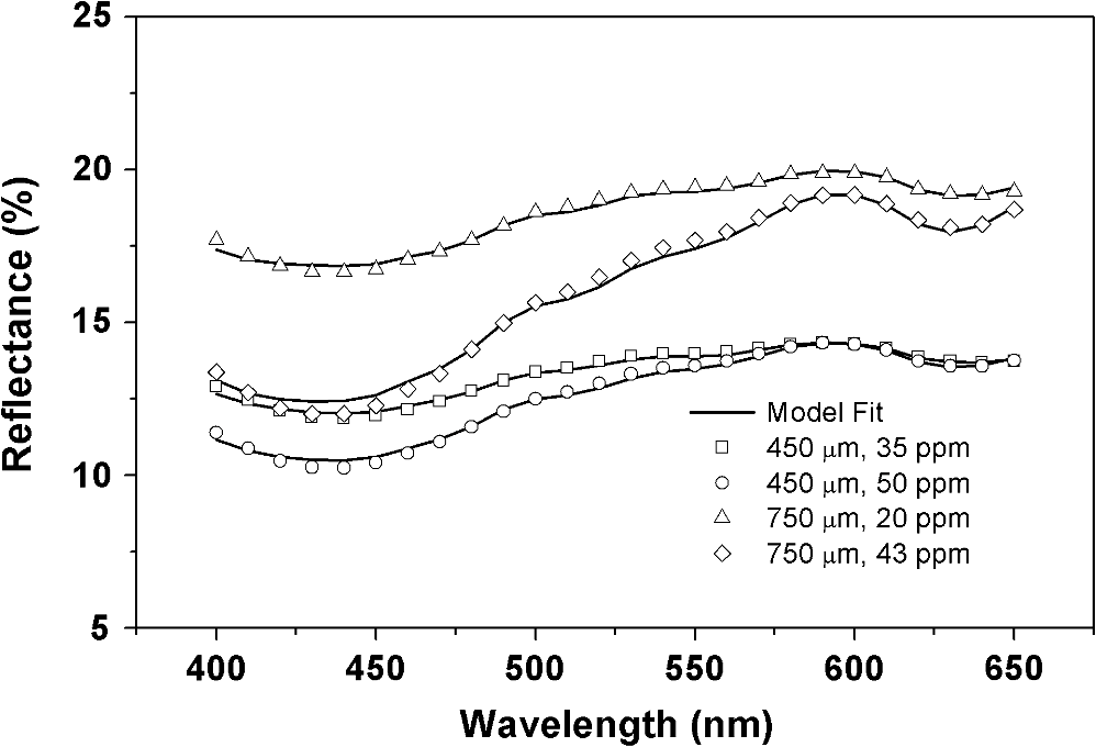 Table 1Actual and extracted values of dye concentrations and phantom thicknesses along with error in prediction.
The dye concentration and phantom thickness were used as fitting parameters in the model and simultaneously varied to obtain the best fit to the reflectance curves shown in Fig. 8. 4.DiscussionIn most biological tissues, diffusion approximation is valid only in the wavelength region between 600 and 900 nm where scattering is larger than the absorption. Sometimes, even in the highly scattering regime, collimated incident beams do not become diffusive if the tissues studied are very thin. For example, the epidermis of skin is only about 50 to 100 μm thick, where absorption is more than the scattering in the 400- to 600-nm region and the transport is not diffusive.22 Also, in most of the measurement geometries, light is not diffusive, especially near the tissue boundaries. Hence, most of the diffusion theory-based models relating K-M coefficients to the radiative transfer coefficients have limited practical applicability. In this paper, a simple and practical empirical relation is developed between K-M and radiative transfer coefficients for a collimated incident beam (either narrow or broad), which is typical of most of the measurement geometries, for example, integrating sphere-based reflectance measurements. The relations developed are also shown be to be applicable in the nondiffusive regimes where absorption is larger than scattering. The empirical relation in this model was derived by measuring the total reflectance and total transmittance of the tissue phantoms. One of the key challenges in measuring total reflectance using integrating sphere geometry is the collection of all scattered light with minimal loss. An incident beam diverges within the sample due to scattering, and the reflected beam size is always larger than the incident beam. In the case of thin or low-scattering samples, the beam divergence will be minimal and hence the error in reflectance measurement will be small. For thick and highly scattering samples, the divergence of the beam will be higher. The error in reflectance measurement can increase, thereby decreasing the accuracy of this method. This error can be reduced by the use of integrating spheres with a larger aperture size. In the current study, we find that, in the typical tissue scattering regime (), the empirical relations seem to works well with less than 10% error in solving the inverse problem. Most of the existing empirical models for studying tissue reflectance are applicable only to homogeneous tissues and cannot be extended to inhomogeneous or layered tissues. Many authors have developed multi-layer models to describe reflection from inhomogeneous tissues. Similarly, since the K-M theory provides analytical expression of both transmittance and reflectance of the diffusive layers, it can be extended to describe layered tissues also. If and are the reflectance and transmittance of the different layers that can be calculated using Eq. (1) and the empirical relations shown in Eqs. (3) and (4), then the total reflectance and transmittance of the layers can be calculated in the following way. If is the light flux traveling in the forward direction and is the light traveling in the reverse direction from the th layer, respectively, then we can write The total reflectance and total transmittance . With and as boundary conditions, these equations can be solved self-consistently to obtain the values of . For example, for a two-layer tissue, solving Eq. (5) self-consistently results in the following relation: Similarly, for a tissue with inhomogeneous distribution of chromophores, a multilayer model can be built by discretizing the layers with a distribution of absorption and scattering coefficients. However, like any other multilayer model, the current model also will have multiple parameters describing the layers, and hence one needs to solve the inverse problem of obtaining these parameters from the measured reflectance. 5.ConclusionsIn summary, by measuring total transmittance and total reflectance of tissue phantoms with varying optical parameters, we have obtained empirical relations between K-M coefficients and the radiative transport coefficients for integrating sphere-based spectrophotometers that use uniform, nondiffusive incident beams. We have shown that the K-M scattering coefficient depends only on reduced scattering coefficient, while the K-M absorption coefficient depends on both the absorption and reduced scattering coefficients of the radiative transfer theory. We have also shown that these empirical relations are valid in both the diffusive and nondiffusive regimes. Using these relations, we could predict reflectance spectra of tissues within an error of 6%. The empirical relations were used to predict the chromophore concentration and the tissue thickness simultaneously by solving the inverse problem from the measured reflectance. The error in the prediction of chromophore concentration and tissue thickness was less than 10%. ReferencesB. W. Murphyet al.,
“Toward the discrimination of early melanoma from common and dysplastic nevus using fiber optic diffuse reflectance spectroscopy,”
J. Biomed. Opt., 10
(6), 064020
(2005). http://dx.doi.org/10.1117/1.2135799 JBOPFO 1083-3668 Google Scholar
F. Koeniget al.,
“Spectroscopic measurement of diffuse reflectance for enhanced detection of bladder carcinoma,”
Urology, 51
(2), 342
–345
(1998). http://dx.doi.org/10.1016/S0090-4295(97)00612-2 0090-4295 Google Scholar
I. J. Bigioet al.,
“Diagnosis of breast cancer using elastic-scattering spectroscopy: preliminary clinical results,”
J. Biomed. Opt., 5
(2), 221
–228
(2000). http://dx.doi.org/10.1117/1.429990 JBOPFO 1083-3668 Google Scholar
J. R. Mourantet al.,
“Spectroscopic diagnosis of bladder cancer with elastic light scattering,”
Lasers Surg. Med., 17
(4), 350
–357
(1995). http://dx.doi.org/10.1002/(ISSN)1096-9101 LSMEDI 0196-8092 Google Scholar
A. Ishimaru, Wave Propagation and Scattering in Random Media, Academic Press, New York
(1978). Google Scholar
K. M. CaseP. F. Zweifel, Linear Transport Theory, Addison-Wesley, Reading, PA
(1967). Google Scholar
B. C. WilsonG. Adam,
“A Monte Carlo model for the absorption and flux distributions of light in tissue,”
Med. Phys., 10
(6), 824
–830
(1983). http://dx.doi.org/10.1118/1.595361 MPHYA6 0094-2405 Google Scholar
S. A. Prahlet al.,
“A Monte Carlo model of light propagation in tissue,”
Proc. SPIE, IS 5 102
–111
(1989). PSISDG 0277-786X Google Scholar
G. MantisG. Zonios,
“Simple two-layer reflectance model for biological tissue applications,”
Appl. Opt., 48
(18), 3490
–3496
(2009). http://dx.doi.org/10.1364/AO.48.003490 APOPAI 0003-6935 Google Scholar
R. ReifO. A’AmarI. J. Bigio,
“Analytical model of light reflectance for extraction of the optical properties in small volumes of turbid media,”
Appl. Opt., 46
(29), 7317
–7318
(2007). http://dx.doi.org/10.1364/AO.46.007317 APOPAI 0003-6935 Google Scholar
T. J. FarrellM. S. PattersonB. C. Wilson,
“A diffusion theory model of spatially resolved, steady-state diffuse reflectance for the non-invasive determination of tissue optical properties in vivo,”
Med. Phys., 19
(4), 879
–888
(1992). http://dx.doi.org/10.1118/1.596777 MPHYA6 0094-2405 Google Scholar
A. KienleM. S. Patterson,
“Improved solutions of the steady-state and the time resolved diffusion equations for reflectance from a semi-infinite turbid medium,”
J. Opt. Soc. Am. A, 14
(1), 246
–254
(1997). http://dx.doi.org/10.1364/JOSAA.14.000246 JOAOD6 0740-3232 Google Scholar
P. KubelkaF. Munk,
“Ein Beitrag zur Optik der Farbanstriche,”
Zeits. f. tech. Physik, 12 593
–601
(1931). Google Scholar
K. Klier,
“Absorption and scattering in plane parallel turbid media,”
J. Opt. Soc. Am., 62
(7), 882
–885
(1972). http://dx.doi.org/10.1364/JOSA.62.000882 JOSAAH 0030-3941 Google Scholar
M. J. C. van GemertW. M. Star,
“Relations between the Kubelka–Munk and transport equation models for anisotropic scattering,”
Lasers Life Sci., 1 287
–298
(1987). Google Scholar
B. J. Brinkworth,
“Interpretation of the Kubelka–Munk coefficients in reflection theory,”
Appl. Opt., 11
(6), 1434
–1435
(1972). http://dx.doi.org/10.1364/AO.11.001434 APOPAI 0003-6935 Google Scholar
L. F. Gate,
“Comparison of the photon diffusion model and Kubelka–Munk equation with the exact solution of the radiative transport equation,”
Appl. Opt., 13
(2), 236
–238
(1974). http://dx.doi.org/10.1364/AO.13.000236 APOPAI 0003-6935 Google Scholar
J. T. Atkins,
“Absorption and scattering of light in turbid media,”
University of Delaware,
(1965). Google Scholar
J. Reichman,
“Determination of absorption and scattering coefficients for non-homogeneous media. 1: theory,”
Appl. Opt., 12
(8), 1811
–1815
(1973). http://dx.doi.org/10.1364/AO.12.001811 APOPAI 0003-6935 Google Scholar
S. N. Thennadil,
“Relationship between the Kubelka–Munk scattering and radiative transfer coefficients,”
J. Opt. Soc. Amer. A., 25
(7), 1480
–1485
(2008). http://dx.doi.org/10.1364/JOSAA.25.001480 JOAOD6 1084-7529 Google Scholar
Philip Laven,
“A computer program for scattering of light from a sphere using Mie theory & the Debye series,”
(2012) http://www.philiplaven.com/mieplot.htm ( October ). 2012). Google Scholar
T. IgarashiK. NishinoS. Nayar,
“The appearance of human skin,”
(2005). Google Scholar
|
||||||||||||||||||||||||||||||||||||