|
|
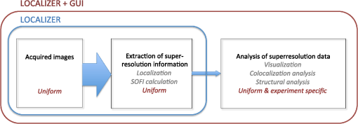
1.IntroductionAny biological system adapts and survives through a multitude of interactions and reactions. Fluorescence imaging has become one of the major avenues for analyzing the many molecular and cellular events underlying these processes. However, even though most fluorophores can be used as molecular labels, the direct observation of the molecular length scale, using optical microscopy, is precluded due to the diffraction of light. To circumvent this inherent limit, a number of techniques have recently been developed that provide far-field fluorescence microscopy with a fundamentally unlimited spatial resolution. These “superresolution” approaches are capable of painting the picture of life with an unprecedented level of detail.1 Techniques such as the localization microscopies [e.g., photo-activation localization microscopy (PALM),2,3 stochastic optical reconstruction microscopy (STORM),4 and ground-state depletion imaging (GSDIM)5] and fluctuation microscopy [superresolution optical fluctuation imaging (SOFI)6 and photochromic stochastic optical fluctuation imaging (pcSOFI)7] can be performed with what is essentially a standard optical microscope. Despite this apparent accessibility and increasing availability of commercial implementations, superresolution microscopy remains a technology in development, rather than one being routinely applied. A significant consideration for any group planning a superresolution experiment is the data analysis. There are a handful of good programs and algorithms that are freely available for superresolution analysis. However, to our knowledge, none of the existing packages offer a complete analytical environment for both localization analysis and fluctuation analysis, with a choice of algorithms for each. Furthermore, to the best of our knowledge, there is no existing software that provides a modular, customizable program that can be readily tailored to the needs of the individual user. Here, we present Localizer,8 an open-source program for implementing superresolution localization analysis (PALM, STORM, dSTORM) and fluctuation analysis (SOFI/pcSOFI). Localizer has been developed to serve two purposes: to be a robust, accurate and precise tool for superresolution microscopy and also to be an adaptable and maintainable platform for those labs that need to customize their analysis or aim to develop new applications for superresolution microscopy. 2.Results and DiscussionExperiments based on localization or fluctuations can fundamentally be divided into three distinct phases tailored to the specific question that the user aims to address (Fig. 1):
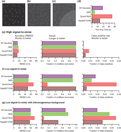
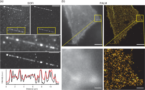
Fig. 1Schematic overview of a superresolution localization or SOFI analysis. The acquired images are first reduced to a smaller dataset by performing fluorophore localization or cumulant analysis. This reduced dataset is then used for further experiment-specific processing. The size of the arrows denotes the relative amount of data that is transferred. The exterior boxes indicate the capabilities available in Localizer itself and in the combination of Localizer and the included Igor Pro graphical interface.  Step 1 necessitates the recording of hundreds or thousands of fluorescence images using standard wide-field fluorescence microscopy, for both stochastic optical fluctuation imaging and the superresolution techniques based on the localization of individual emitters. Localizer has been designed to rapidly and accurately implement Step 2 for both the localization and fluctuation microscopies, directly processing long input data sequences (movies) according to user-specified settings and outputting a set of localized positions or one or more computed images. Localizer can be run from within Igor Pro (for which an intuitive graphical user interface has been developed) or Matlab, such that Step 3, the post-processing and subsequent analysis of data, can be readily achieved from within the same software environment. As an alternative, Localizer can be run as a stand-alone program from the command line. In addition, the software was designed to be portable across platforms or software packages. By virtue of its crucial role in the analytical process, the software used to process superresolution data can have a significant impact on the conclusions that can be drawn from an experiment: Inaccuracies, artifacts, or suboptimal performance will effectively contaminate the downstream analyses and images. Hence, of primary concern to us, was the development of a robust analytical tool. To evaluate the performance and accuracy of Localizer, we set out to compare the program with other software packages. However, this comparison was impossible for the SOFI analysis since we were unable to find other freely available implementations. For localization microscopy we compared the program with two freely available packages: rapidSTORM,9 a stand-alone program for Windows and Linux, and QuickPALM,10 a plug-in for the ImageJ microscopy analysis platform. Since the absolute positions of the emitters are unknown in actual imaging data, we generated three simulated datasets, in which we attempted to model the imaging process as closely as possible. This includes the use of a non-Gaussian point spread function calculated using vectorial optics, non-correlated background photons, as well as the effects of electron-multiplied CCD (EMCCD) amplification and readout. We considered three different scenarios: an “easy” dataset, with very bright emitters on a uniform background, a dataset generated under conditions of low signal-to-noise and uniform background, and a dataset with low signal-to-noise and a non-uniform background [Fig. 2(a)–2(c)]. To illustrate the modularity of Localizer, we ran the software using two localization algorithms: the default, involving non-weighted least-squares fitting of a Gaussian function, and Gaussian mask estimation (GME).11 In all cases, we performed a best-effort optimization of the analysis in each program, without consulting the actual emitter locations. Fig. 2Comparison of Localizer with other freely-available analysis packages. (a)–(c) Example simulated images for high signal-to-noise (a), low signal-to-noise (b), and low signal-to-noise with non-uniform background (c). (d) Time required to analyze 500 images with high signal-to-noise. (e)–(g) Performance of each software package under the conditions shown in (a)–(c) (RMSD refers to the root mean square localization deviation). Two different localization algorithms implemented in Localizer were included (2D Gauss fitting and GME) in the comparison.  We quantified the performance of each analysis using three different parameters: the accuracy, the recall (the fraction of simulated emitters that was recovered), and the error rate (the fraction of reported emitters that was spurious). Finally, we also compared the time required to perform the complete analysis. The results are summarized in Fig. 2. Predictably, we found that all programs performed well on the “easy” dataset [Fig. 2(e)]. QuickPALM performed slightly worse than both Localizer and rapidSTORM in every respect, while rapidSTORM provided a slightly better localization accuracy, possibly due to its default of fixed-width Gaussian fitting. Under conditions of low signal-to-noise Fig. 2(f)], QuickPALM effectively fails to provide useful data, though rapidSTORM and Localizer continue to yield comparable performance. However, for very low signal-to-noise ratios we found that rapidSTORM provided somewhat better segmentation compared to the default GLRT algorithm12 in Localizer. Finally, under conditions of low signal-to-noise and non-uniform background [Fig. 2(g)], which can occur when imaging different cellular compartments, such as the cytosol and nucleus, we find that Localizer yields better results. Here, the modularity of Localizer also plays an important role in ensuring the software’s performance. Whilst more emitters were localized using the GME fitting than the default non-linear least-squares fitting, the GME fitting also results in a significantly greater fraction of false positives (around 10%) compared with the default fitting (negligible). Here, the data analysis is sufficiently demanding that optimization of not only the fitting parameters, but the fitting algorithm is necessary to obtain a reliable superresolution image. Figure 3 shows some examples of the output of Localizer, produced using both fluctuation and localization microscopy. Figure 3(a) shows SOFI and average images taken from a movie 400 frames in length of fragments of a T7 phage DNA molecule stained using a DNA intercalating dye and imaged under conditions that allow reversible binding of the dye (YOYO-1).13 Figure 3(b) shows PALM data acquired on a fixed HeLa cell stained using Dendra2 targeted to the plasma membrane using the LyN targeting motif. Additional examples of images resulting from the application of Localizer can be found in the literature.7,14–16 Fig. 3Example of superresolution data processed and visualized using Localizer. (a) Average (left) and superresolution SOFI (right) images of fragments (20 to 25 kb in length) of T7 phage DNA molecules stained with YOYO-1 dye under conditions that promote reversible binding of the YOYO-1. Movies of 400 frames in length were processed using Localizer to derive both images. The lower images show an expansion of the region highlighted in yellow, and a line profile along the length of the DNA molecule at the bottom. Scale bars denote 5 and 2 μm, respectively. (b) Conventional (left) and PALM (right) image of Lyn-Dendra2 targeted to the plasma membrane of a fixed HeLa cell. The scale bars correspond to 5 μm and 500 nm.  One of our main concerns in developing Localizer was to ensure that it is analytically flexible yet retains the properties of being modifiable and expandable with minimal effort. Such properties are critical in superresolution microscopy, where the field of research is at an evolutionary stage, and the algorithms that form the foundations of these techniques are still under development. Furthermore, as with standard fluorescence microscopy, the range of applications and diversity of samples can be vast. As we have shown with our simple, simulated data, the quality of the raw data that is input to the analysis software can significantly impact the ability of a given program or algorithm to produce a reliable, or complete, superresolution image. For example, consistent with previous reports,17 we have shown that there is significant variability in the performance of the localization algorithms. The optimal choice or combination of choices of algorithms depends not only on their intrinsic abilities but also on the measurement conditions and desired analysis. Thus, in our opinion, the modularity of Localizer is not merely an “added extra” but rather a critical component of any software that is to be widely employed for producing superresolution images. As written, Localizer offers a number of options for both optimal segmentation (particle detection) and optimal particle localization. This choice of thresholding and localization algorithms can be either made within the Igor Pro GUI or by changing a single argument in the Matlab, Igor Pro, or other bridges. Furthermore, since the Localizer code is composed as a series of modules, new algorithms can be readily incorporated in the program without modifying the existing code. A tutorial on how to expand or adapt Localizer is supplied along with the source code. Localizer is written in the C++ programming language, which allows it to combine high performance with the ability to run on any computer platform, without requiring specific software. Creating new bindings (to run Localizer from within software other than Matlab or Igor Pro) requires only the addition of small code segments specific to that environment, and does not require modifications to the core code of Localizer, which is shared across all implementations. Like the core program itself, the bridges and interface are all available as free and open source software (GPL license). 3.ConclusionIn conclusion, we have presented Localizer, a software package for accurate and fast analysis of superresolution measurements, including localization (PALM/STORM/GSDIM) and SOFI/pcSOFI microscopy. Localizer delivers high accuracy and performance and comes with a fully featured and easy-to-use graphical interface but is also designed to be integrated in higher-level analysis environments. Due to its modular design, Localizer can be readily extended with new algorithms as they become available, while maintaining the same interface and performance. We provide front-ends for running Localizer from Igor Pro, Matlab, or as a stand-alone program. We find that Localizer performs favorably when compared with two existing superresolution packages, QuickPALM and RapidSTORM, and to our knowledge is the only freely available implementation of SOFI/pcSOFI microscopy. By dramatically improving the analysis performance and ensuring the easy addition of current and future enhancements, Localizer strongly improves the usability of superresolution imaging in a variety of biomedical settings. AcknowledgmentsPeter Dedecker thanks the Research-Foundation Flanders (FWO-Vlaanderen) for a postdoctoral fellowship and travel grant. This work was supported by an NIH Director’s Pioneer Award DP1 OD006419 and NIH grants R01 DK073368 and DP1CA174423 (to J. Z.). ReferencesS. W. Hell,
“Far-field optical nanoscopy,”
Science, 316
(5828), 1153
–1158
(2007). http://dx.doi.org/10.1126/science.1137395 SCIEAS 0036-8075 Google Scholar
E. Betziget al.,
“Imaging intracellular fluorescent proteins at nanometer resolution,”
Science, 313
(5793), 1642
–1645
(2006). http://dx.doi.org/10.1126/science.1127344 SCIEAS 0036-8075 Google Scholar
S. T. HessT. P. K. GirirajanM. D. Mason,
“Ultra-high resolution imaging by fluorescence photoactivation localization microscopy,”
Biophys. J., 91
(11), 4258
–4272
(2006). http://dx.doi.org/10.1529/biophysj.106.091116 BIOJAU 0006-3495 Google Scholar
M. J. RustM. BatesX. Zhuang,
“Sub-diffraction-limit imaging by stochastic optical reconstruction microscopy (STORM),”
Nat. Methods, 3
(10), 793
–795
(2006). http://dx.doi.org/10.1038/nmeth929 1548-7091 Google Scholar
J. Föllinget al.,
“Fluorescence nanoscopy by ground-state depletion and single-molecule return,”
Nat. Methods, 5
(11), 943
–945
(2008). http://dx.doi.org/10.1038/nmeth.1257 1548-7091 Google Scholar
T. Dertingeret al.,
“Fast, background-free, 3D super-resolution optical fluctuation imaging (SOFI),”
Proc. Natl. Acad. Sci. U.S.A., 106
(52), 22287
–22292
(2009). http://dx.doi.org/10.1073/pnas.0907866106 PNASA6 0027-8424 Google Scholar
P. Dedeckeret al.,
“Widely accessible method for superresolution fluorescence imaging of living systems,”
Proc. Natl. Acad. Sci. U.S.A., 109
(27), 10909
–10914
(2012). http://dx.doi.org/10.1073/pnas.1204917109 PNASA6 0027-8424 Google Scholar
S. Wolteret al.,
“Real-time computation of subdiffraction-resolution fluorescence images,”
J. Microsc. Oxford, 237
(1), 12
–22
(2010). http://dx.doi.org/10.1111/(ISSN)1365-2818 JMICAR 0022-2720 Google Scholar
R. Henriqueset al.,
“QuickPALM: 3D real-time photoactivation nanoscopy image processing in Image,”
Nat. Methods, 7
(5), 339
–340
(2010). http://dx.doi.org/10.1038/nmeth0510-339 1548-7091 Google Scholar
R. E. ThompsonD. R. LarsonW. W. Webb,
“Precise nanometer localization analysis for individual fluorescent probes,”
Biophys. J., 82
(5), 2775
–2783
(2002). http://dx.doi.org/10.1016/S0006-3495(02)75618-X BIOJAU 0006-3495 Google Scholar
A. Sergeet al.,
“Dynamic multiple-target tracing to probe spatiotemporal cartography of cell membranes,”
Nat. Methods, 5
(8), 687
–694
(2008). http://dx.doi.org/10.1038/nmeth.1233 1548-7091 Google Scholar
I. Schoenet al.,
“Binding-activated localization microscopy of DNA structures,”
Nano. Lett., 11
(9), 4008
–4011
(2011). http://dx.doi.org/10.1021/nl2025954 NALEFD 1530-6984 Google Scholar
M. J. Roeffaerset al.,
“Super-resolution reactivity mapping of nanostructured catalyst particles,”
Angewandte Chemie International Edition, 48
(49), 9285
–9289
(2009). http://dx.doi.org/10.1002/anie.v48:49 ACIEAY 0570-0833 Google Scholar
S. A. Ribeiroet al.,
“A super-resolution map of the vertebrate kinetochore,”
Proc. Natl. Acad. Sci., 107
(23), 10484
–10489
(2010). http://dx.doi.org/10.1073/pnas.1002325107 1091-6490 Google Scholar
R. K. Neelyet al.,
“DNA fluorocode: a single molecule, optical map of DNA with nanometre resolution,”
Chem. Sci., 1
(4), 453
(2010). http://dx.doi.org/10.1039/c0sc00277a CESCAC 0009-2509 Google Scholar
K. I. Mortensenet al.,
“Optimized localization analysis for single-molecule tracking and super-resolution microscopy,”
Nat. Methods, 7
(5), 377
–381
(2010). http://dx.doi.org/10.1038/nmeth.1447 1548-7091 Google Scholar
|

