|
|
1.IntroductionBone fracture healing is a complex biological process that involves the spatial and temporal interaction of various cell types with large numbers of genes and an extracellular matrix.1 The process of fracture healing can be divided into several stages such as immediate injury response, inflammatory phase, intramembranous and endochondral ossification and, finally, bone remodeling.2 Each step presents distinct histological features but they all depend on the expression of cell-signaling molecules, synthesis of genes, deposition of extracellular matrix, and bone cell proliferation and differentiation.1,2 Moreover, it is well known that each phase shows distinct histological features, with the marked interaction of osteoblasts and osteoclasts cells.3 Osteoblasts have an important role in bone formation, being responsible by forming bone tissue, and their activity is influenced by Runx-2, bone morphogenetic proteins (BMPs), alkaline phosphatase (ALP), type I collagen, proteoglycan, bone sialoprotein, and osteocalcin (OC).4–9 In general, bone tissue has the ability of healing by itself. However, under critical conditions as for example, in larger bone defects and fractures with poor vascularization, a delay in the process of healing or even a nonunion can happen.10 Within this context, innovative clinical approaches to accelerate bone metabolism and to repair damage to bone tissue are being developed.11 One promising treatment method is the use of low-level laser therapy (LLLT).11 Several studies suggest that LLLT can stimulate osteogenesis at the site of fracture, promoting a higher deposition of bone mass and accelerating the consolidation process.11–13 The effects of LLLT in biological tissues can be explained by the photochemical theory, which offers an explanation for the sensitivity of cells to electromagnetic light.14 Laser irradiation is absorbed by photoreceptors located in the cells, promoting changes in morphology molecular drum and thereafter. This starts a series of modifications in cellular processes, with the increase in cell signaling and excitation of components of the mitochondrial respiratory chair.15,16 The stimulation of the respiratory chain alters the redox potential of the cytoplasm and mitochondria, accelerating electron exchange and consequently increasing ATP production. Therefore, one can observe an increase in cellular metabolism that leads to a stimulation of DNA and RNA expression, proteins, and enzymes synthesis.17 Stein et al.18 investigated the effects of LLLT (670 nm, , 400 mW) on human osteoblast-like cells and observed a significant increase in mRNA expression of ALP after 48 and 72 h in the irradiated group as well as an upregulation in the mRNA expression of osteopontin after 24 h. Moreover, Fávaro-Pipi et al.19 evaluated the effects of LLLT (830 nm, 30 mW, ) on the synthesis of genes in a tibial bone defect model. The authors demonstrated a higher expression of BMP-4 at day 13 post-surgery and an upregulation of mRNA of BMP4, ALP and Runx-2 at day 25 after surgery.19 Although the effects of LLLT have been well described, the molecular and cellular changes induced by this treatment on bone tissue are not fully understood. Furthermore, there is a lack in the literature describing the mechanisms by which laser acts in the initial periods in LLLT repair, since most of studies have evaluated it effects on intermediate and late periods of bone healing.1,12,13 Therefore, the hypothesis of this study is that LLLT is capable of improving bone repair in the initial periods of consolidation by activating the expression of genes related to bone cell proliferation and differentiation. Considering the stimulatory effects of LLLT on bone regeneration after an injury, the aim of the present study was to evaluate the morphological changes of bone and to measure the temporal pattern of the expression of osteogenic genes after LLLT during the initial process of bone healing. We used histopathology and quantitative real time polymerase chain reaction (RT-qPCR) analysis to evaluate the effects of laser therapy irradiation in bone defects in tibias of rats at 12 h, 36 h, 3 d and 5 d postsurgery. 2.MethodsThis study was approved by the Committee of Ethics in Animal Research at the Federal University of São Paulo, Brazil (0975/10). A total of 64 male Wistar rats (weighing , 12 to 13 weeks) were distributed randomly into two groups: control or laser irradiated. They were maintained under controlled temperature (), light-dark periods of 12 h, and with free access to water and a commercial diet. All animal handling and surgical procedures were strictly conducted according the Guiding Principles for the Use of Laboratory Animals. As described below, noncritical size bone defects were performed on both tibias. Rats were sacrificed at 12 h, 36 h, 3 d and 5 d after surgery. 2.1.SurgeryNoncritical size bone defects (2.5 mm diameter) were surgically created at the upper third of the tibia (10 mm distal of the knee joint). Surgery was performed under sterile conditions and general anesthesia induced by intraperitoneal injection of xilazin (Anasedan; Vetbrands, São Paulo, Brazil) and ketamin (Dopalen; Vetbrands, São Paulo, Brazil). After antiasepsia of the skin, the medial compartment of the tibia was exposed through a longitudinal incision on the shaved skin and muscle tissue. A standardized 2.5 mm diameter bone defect was created using a motorized drill (Dentsply, Maillefer Instruments SA, Ballaigues, Suíça), 12.356 rpm, under copious irrigation with saline solution. The cutaneous flap was replaced and sutured with resorbable polyglactin, and the antisepsis was performed with povidone iodine. The animals received analgesia (i.m., buprenorphine (Temgesic; Reckitt Benckiser Health Care Limited, Schering Plough, Hoddesdon, United Kingdom) and were returned to their cages. The health status of the rats was monitored daily.12,19 2.2.TreatmentsThe treatments, which started immediately post-surgery, were performed for one (12 h), two (36 h), three (3 d) or five (5 d) sessions, with an interval of 24 h. A low-energy gallium–aluminum–arsenide (Ga-Al-As), laser 830 nm, 30 mW, continuous wavelength, , , 1 min and 34 s, 2.8 J, (Tera laser, DMC Equipamentos, São Carlos, SP, Brazil) was used. The parameters were specified based on the equipment specification of the manufacturer. The irradiation was performed at one point, above the area of the injury, through the punctual contact technique. On 12 h, 36 h, 3 d and 5 d post-injury, rats were sacrificed individually by carbon dioxide asphyxia. Both tibias were removed for analysis. 2.3.Histopathological AnalysisFor the qualitative histopathological analysis, the right tibia was used. They were removed, fixed in 10% buffer formalin (Merck, Darmstadt, Germany) for 48 h, decalcified in 4% EDTA (Merck, Darmstadt, Germany), and embedded in paraffin blocks. Five-micrometer slices were obtained in serial sections in the longitudinal direction and stained with hematoxylin and eosin (H.E stain, Merck). A descriptive qualitative histopathological evaluation of the total area of the bone defect was performed by a pathologist (blinded to the treatment) under a light microscope (Olympus, Optical Co. Ltd, Tokyo, Japan) at magnification. Any changes in the bone defect, such as presence of woven bone, bone marrow, inflammatory process, granulation tissue, or even tissues undergoing hyperplastic, metaplastic, and/or dysplastic transformation were investigated per animal. 2.4.Total RNA Isolation and Real-Time Polymerase Chain ReactionImmediately postmortem, left tibias were dissected (periosteum intact) and rapidly frozen in liquid nitrogen. The region of the callus was stored () until analysis by RT-qPCR. Total RNA was isolated using standard protocols. Trizol reagent (1 ml, Invitrogen, Carlsbad, California) was added to the sample and allowed to thaw. The mixture was transferred to a polypropylene tube and incubated (room temperature, 5 m). Chloroform (0.2 ml, Sigma, St. Louis, Missouri) was added, mixed vigorously, and the mixture was transferred to a 2 ml tube (Eppendorf, Hamburg, Germany) and centrifuged (28°C, 15 m). The nucleic acid phase was decanted and an equal volume of RNase-free 70% ethanol was added. Potential DNA contamination was removed by RNase-free DNase I (Invitrogen). RNA integrity was verified by RNA gel electrophoresis and spectrophotometry. Four genes of interest were selected representing processes associated with osteogenesis (Table 1). For each gene, rat-specific primers were designed for real-time PCR around exon junctions where possible. All real-time primers were initially tested against standards and a standard curve was generated. First strand cDNA was synthesized (M-MLV RT, Invitrogen) from total RNA (1 mg). RT-qPCR reactions were carried out at 50 ml total volume. Following an initial denaturing step for each primer (Table 1), the genes of interest were amplified through 40 cycles (Rotor-Gene, R 3000, Robert Research, Mortlake, Australia). Gene amplification was measured by SYBR green (Applied Biosystems, Carlsbad, California) fluorescence during the annealing/elongation phase. All samples were run in duplicate and the average was used for further analysis. Measures of real-time PCR cycle to threshold were normalized to the expression of ribosomal protein S18 (RPS18) for each tibia. For comparison between experimental groups, RPS18 normalized expression from each tibia was divided by the normalized gene expression from the control group to obtain a fold increase in gene expression of LLLT group and control group in 12 h, 36 h, 3 d and 5 d. Table 1Primers and indicated annealing temperatures for each gene analyzed.
Note: OC, osteocalcin; ALP, alkaline phosphatase; Runx -2, transcriptional factor. 2.5.Statistical AnalysisData are expressed as the error of the mean (SEM). The normality of all variables’ distribution was verified using Shapiro–Wilk’s W test. Two-way ANOVA was used to assess the effect of displacement level and time. Post hoc, multiple comparisons were made using Tukey’s protected least significant difference tests with statistical significance defined as . 3.ResultsFrom the 80 animals available for this study, four animals were lost due to an anesthesia-induced respiratory depression. The remaining animals recovered uneventfully from the surgical procedure and remained in good health. No signs of macroscopic inflammation or adverse tissue responses were observed during the experimental period. At the end of the experiment, 152 tibias were retrieved and used for both analyses (histopathology and RT-qPCR). For histopathological evaluation, 80 tibias were used, however four tibias were excluded for animals that died during surgery. In addition, four tibias were excluded from analysis during histological processing (Table 2). Furthermore, a total of 72 tibias were retrieved, of which 68 of them were included for histopathology analysis. For RT-qPCR, five animals per group were used in a total of 40 tibias. Table 2Number of tibias, retrieved and used for histological analyses.
3.1.Histopathological AnalysisTwelve hours after surgery, for the animals in the control group, the edges of the defect were visible and a moderate amount inflammatory infiltrate, with polymorphonuclear cells (mainly represented by neutrophils) were observed [Fig. 1(a)]. In the LLLT group, the histopathological analysis revealed an intense amount of inflammatory infiltration, with a higher presence of polymorphonuclear cells (neutrophils) when compared to control [Fig. 1(b)]. Fig. 1Bone defects (a) control group 12 h; (b) LLLT group 12 h; (c) control group 36 h; (d) LLLT group 36 h; (e) control group three days; (f) LLLT group three days; (g) control group five days; (h) LLLT group five days. (*) inflammatory infiltrate, (⋆) granulation tissue, (→) early woven bone formation. H.E.(200×). 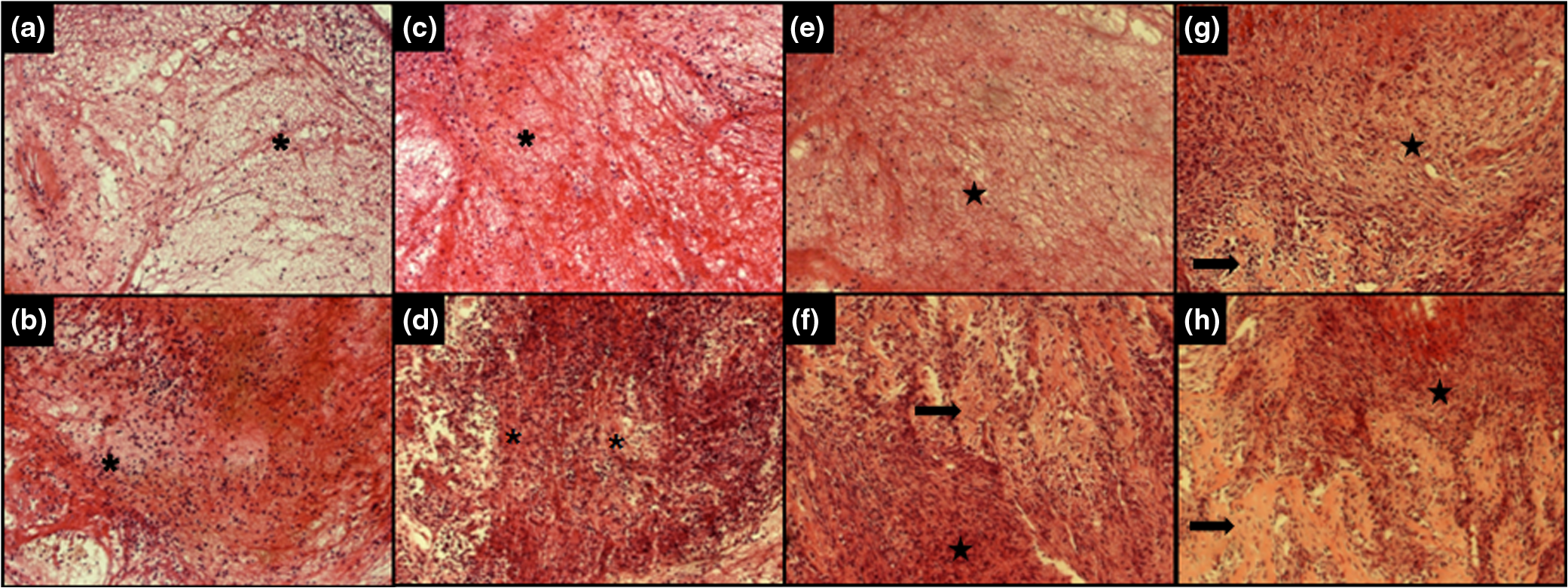 After 36 h, the control group presented an intense inflammatory infiltration, with polymorphonuclear cells [Fig. 1(c)]. In the laser-irradiated animals, a lower inflammatory response was observed when compared to control [Fig. 1(d)]. In addition, on day three, a decrease in the inflammatory process and increased granulation tissue was observed in the control group [Fig. 1(e)]. Also, the irradiated animals displayed a higher amount of granulation tissue, new bone tissue, and neoangiogenesis, which corresponds to more advanced repair compared to controls [Fig. 1(f)]. After five days, histological analysis of the control group [Fig. 1(g)] revealed the presence of granulation tissue and newly formed bone on half of the animals tested. Figure 1(h) shows that, in the irradiated group, higher amount of granulation tissue and regions of bone remodeling and bone ingrowth were found in all animals, with a better tissue organization compared to the control group. 3.2.mRNA Expression of Runx-2, ALP, and OCFigures 2Fig. 3–4 represent the temporal gene expression of the control and LLLT treated groups.
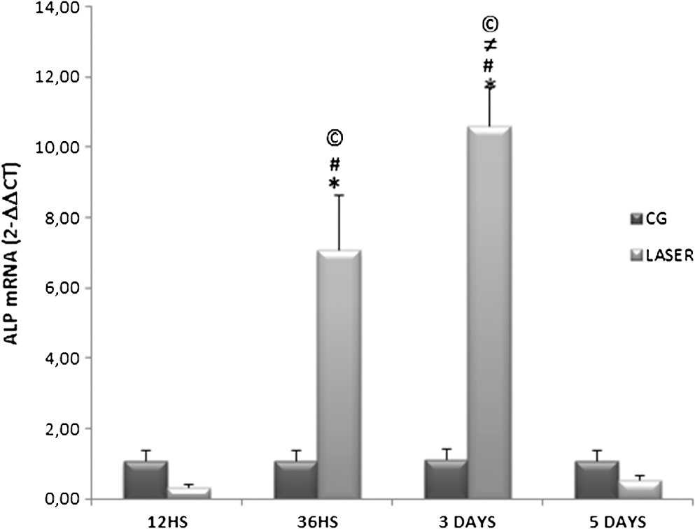
Fig. 2Means and SEM of the changes in the expression of the Runx-2 gene in the control group (CG) and laser-treated group (Laser) measured by the RT-qPCR. ≠ versus laser in 36 h; # versus laser in five days; © versus the respective control group. 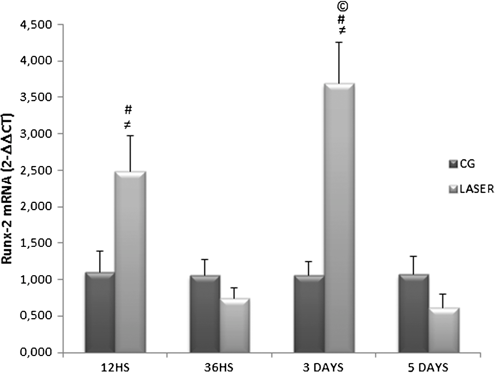 4.DiscussionIn this study the effects of laser therapy on histological modifications and gene expression during the early stage of bone repair were evaluated. It was hypothesized that this therapy would be capable of accelerating tissue healing by the biomodulation of the inflammatory process, stimulation of bone formation, and upregulation of genes related to bone cell differentiation. The main findings showed that laser therapy produced an earlier recruitment of inflammatory cells (neutrophils) as well as a higher amount of granulation tissue and newly formed bone in the irradiated groups. Also, the results of the RT-qPCR showed that laser irradiation produced a significantly increase in mRNA expression of Runx-2, 12 h and three days post-surgery, a significantly upregulation in ALP mRNA expression after 36 h and three days, and a significantly increase in expression of OC mRNA after three and five days. LLLT has been shown to enhance bone healing by increasing mitochondrial respiration and ATP synthesis, increasing of cell proliferation,13,20,21 osteoblasts and fibroblasts cell proliferation and differentiation, stimulation of inflammatory cells, angiogenesis and increased newly formed bone.22,23 Our histopathological analysis revealed that LLLT produced an earlier recruitment of inflammatory cells compared to their respective controls, highlighting its pro-inflammatory effects, which may lead to the earlier resolution of inflammation and anticipation of the other stages of repair.24 Similarly, the higher amount of granulation tissue and newly formed bone in the irradiated groups may be related to the earlier recruitment of osteoprogenitor cells and mature osteoblasts, resulting in higher deposition of newly formed bone. These results corroborate with several studies that showed positive effects of laser irradiation on bone cell proliferation and differentiation.13,21,25 Pretel et al.26 found that LLLT (780 nm, 35 mW, , 1,4 J) modulated the initial inflammatory response and anticipated the resolution of the process to normal conditions at the earlier periods in a bone defect model in rats. Additionally, in vitro and in vivo studies showed that many genes related to bone repair have been shown to have their transcription modified after irradiation with LLLT.27–29 In this study, on the experimental period of 12 h, only Runx-2 mRNA expression was found increased in the LLLT group. Runx-2 is essential for the commitment of early mesenchymal cells to the osteoblasts lineage and differentiation of osteoblasts.30 In addition, Runx-2 controls the differentiation and function of the osteoblast in concert with other factors such as Osterix by regulating the expression of many osteoblast-related genes such as ALP, OC and osteopontin.31 These results corroborate with other studies found in the literature.30–32 For example, Fujimoto et al.32 observed that in osteoblastic cell cultures, LLLT (830 nm, ) promoted an increased expression of Runx-2 12 h and 48 h after irradiation. The higher expression of mRNA Runx-2 could be related to the premature differentiation of osteoblast cells at the site of the injury and could have stimulated the expression of other genes evaluated in this study (ALP and OC). Moreover, RT-qPCR analysis demonstrated that laser irradiation produced an upregulation of the mRNA of ALP at 36 h and three days after the surgery. During the process of bone healing, ALP is an important marker of osteoblast differentiation.33 Our results suggested that the increase of the mRNA of ALP expression in the laser-treated group may have induced the premature recruitment of the osteoblast cells, resulting in a higher deposition of bone matrix. Many authors have demonstrated that LLLT is capable of modulating the expression of the mRNA of ALP at different times.20,28,34 In vitro studies showed that LLLT (904 to 910 nm, 200 mW, ) applied to human osteoblastic cells produced an increased mRNA expression of ALP after 10 days of irradiation.28 However, Khadra et al.27 showed that LLLT (Ga-Al-As, 1, 5 or ) did not show any significant difference in the expression of ALP when applied to cells derived from human mandibular bone. In an in vivo study, Fávaro-Pipi et al.29 observed that laser irradiation (830 nm, , 1.4 J) produced an increase of ALP mRNA expression in a tibial bone defect model in rats, 25 days after the surgery. Interestingly, at the third experimental period evaluated, the mRNA expression of Runx-2 and OC was also significantly increased. Taken as a whole, it appears that this increase in expression of the genes analyzed may suggest that this result is related to differentiation of osteoblasts and newly bone formation observed in the laser-irradiated animals, supporting the hypothesis that laser therapy showed an osteogenic potential. Similarly, OC is a marker of bone formation7 and it is the most abundant noncollagenous proteins in bone tissue.5 In our study, the increased expression of OC mRNA after three and five days in the irradiated animals appears at the same time with the higher amount of newly formed bone in the laser groups observed in the histological analysis. These results corroborate with Stein et al.18 who observed an increase in OC mRNA expression in human osteoblastic cells, 72 h after LLLT application (670 nm, ). The methodology employed in this study has been used by many authors.19,23,35 Some limitations of our work should be pointed out. We investigated the effects of LLLT in the expression of ALP, OC and Runx-2 mRNA during the process of bone healing. It would be interesting to investigate the effects of laser application on the expression of other osteogenic factors that are also involved in bone repair. Also, more quantitative analysis should be included in future researches such as the quantification of inflammatory cells and immunohistochemical analysis. In spite of these limitations, the results of this work highlight the stimulatory effects of laser therapy on bone healing. Summarizing, this study suggests that the increased expression of osteogenic genes may be responsible by the differentiation of osteoblastic cells and stimulation of osteogenesis, which may have culminated in the acceleration of repair in the treated animals (revealed by the histopathological analysis). This work advances in the description of molecular and cellular mechanisms of the effects of LLLT on bone repair process, mainly through the increased expression of genes involved in this process. 5.ConclusionIn conclusion, the present study has shown that LLLT evoked an earlier resolution of the inflammatory process and newly bone formation. In addition, LLLT produced a significant increase of mRNA expression of Runx-2, ALP and OC, which are involved in bone repair. Despite these results, further studies are required to investigate the mechanisms and molecular pathways stimulated by LLLT that culminate in the acceleration of bone healing. ReferencesK. Senaet al.,
“Early gene response to low-intensity pulsed ultrasound in rat osteoblastic cells,”
Ultrasound Med. Biol., 31
(5), 703
–708
(2005). http://dx.doi.org/10.1016/j.ultrasmedbio.2005.01.013 USMBA3 0301-5629 Google Scholar
R. A. Kayalet al.,
“Diabetes causes the accelerated loss of cartilage during fracture repair which is reserved by insulin treatment,”
Bone, 44
(2), 357
–363
(2009). http://dx.doi.org/10.1016/j.bone.2008.10.042 BONEDL 8756-3282 Google Scholar
R. A. Nicolaet al.,
“Effect of low-power GaAlAs laser (660 nm) on bone structure and cell activity, an experimental animal study,”
Lasers Med. Sci., 18
(2), 89
–94
(2003). http://dx.doi.org/10.1007/s10103-003-0260-z LMSCEZ 1435-604X Google Scholar
S. L. Songet al.,
“Temporal expression of proteoglycans in the rat limb during bone healing,”
Gene, 379 92
–100
(2006). http://dx.doi.org/10.1016/j.gene.2006.04.029 GENED6 03781119 Google Scholar
A. Steinet al.,
“Low-level laser irradiation promotes proliferation and differentiation of human osteoblasts in vitro,”
Photomed. Laser Surg., 23
(2), 161
–166
(2005). http://dx.doi.org/10.1089/pho.2005.23.161 PLDHA8 1549-5418 Google Scholar
B. Rathet al.,
“Compressive forces induce osteogenic gene expression in calvarial osteoblast,”
J. Biomech., 41
(5), 1095
–1103
(2008). http://dx.doi.org/10.1016/j.jbiomech.2007.11.024 JBMCB5 0021-9290 Google Scholar
I. Baret al.,
“Molecular imaging of the skeleton: quantitative real-time bioluminescence monitoring gene expression in bone repair and development,”
J. Bone Miner. Res., 18
(3), 570
–578
(2003). http://dx.doi.org/10.1359/jbmr.2003.18.3.570 JBMREJ 0884-0431 Google Scholar
G. R. WohlD. A. TowlerM. J. Silva,
“Stress fracture healing: fatigue loading of the rat ulna induces upregulation in expression of osteogenic and angiogenic genes that mimic the intramembranous portion of fracture repair,”
Bone, 44
(2), 320
–330
(2009). http://dx.doi.org/10.1016/j.bone.2008.09.010 BONEDL 8756-3282 Google Scholar
M. Hadjiargyrouet al.,
“Enhancement of fracture healing by low intensity ultrasound,”
Clin. Orthop. Relat. Res., 355 216
–229
(1998). http://dx.doi.org/10.1097/00003086-199810001-00022 CORTBR 0009-921X Google Scholar
G. Victoriaet al.,
“Bone stimulation for fracture healing: what’s all the fuss?,”
Indian J. Orthop., 43
(2), 117
–120
(1998). http://dx.doi.org/10.4103/0019-5413.50844 INJOAU Google Scholar
S. Hamajimaet al.,
“Effect of low-level laser irradiation on osteoglycin gene expression in osteoblasts,”
Lasers Med. Sci., 18
(2), 78
–82
(2003). http://dx.doi.org/10.1007/s10103-003-0255-9 LMSCEZ 1435-604X Google Scholar
A. L. B. Pinheiroet al.,
“Biomodulatory effects of LLLT on bone regeneration,”
Laser Therapy, 13 73
–79
(2000). http://dx.doi.org/10.5978/islsm.13.73 LATHE5 0898-5901 Google Scholar
Y. A. VladimirovA. N. OsipovG. I. Klebanov,
“Photobiological principles of therapeutic applications of laser radiation,”
Biochemistry, 69
(1), 81
–90
(2004). http://dx.doi.org/10.1023/B:BIRY.0000016356.93968.7e MIRBD9 0144-0578 Google Scholar
T. I. KaruS. F. Kolyakov,
“Exact action spectra of cellular responses relevant to phototherapy,”
Photomed. Laser Surg., 23
(4), 356
–361
(2005). http://dx.doi.org/10.1089/pho.2005.23.355 PLDHA8 1549-5418 Google Scholar
T. I. KaruL. V. PyatibratG. S. Kalendo,
“Cell attachment to extracellular matrices is modulated by pulsed radiation at 820 nm and chemicals that modify the activity of enzymes in the plasma membrane,”
Lasers Surg. Med., 29
(3), 274
–281
(2001). http://dx.doi.org/10.1002/(ISSN)1096-9101 LSMEDI 0196-8092 Google Scholar
S. S. KitchenC. J. Partridge,
“A review of low level laser therapy,”
Physiotherapy, 77
(3), 161
–168
(1991). http://dx.doi.org/10.1016/S0031-9406(10)61694-X 0031-9406 Google Scholar
T. Karu,
“Introduction,”
The Science of Low-Power Laser Therapy, xiii
–xviii Gordon and Breach Science Publishers, Amsterdam
(1998). Google Scholar
A. Steinet al.,
“Initial effects of low-level laser therapy on growth and differentiation of human osteoblast-like cells,”
Wien Klin. Wochenschr., 120
(3–4), 112
–117
(2008). http://dx.doi.org/10.1007/s00508-008-0932-6 0043-5325 Google Scholar
E. Fávaro-Pipiet al.,
“Comparative study of the effects of low-intensity pulsed ultrasound and low-level laser therapy on bone defects in tibias of rats,”
Lasers Med. Sci., 25
(5), 727
–32
(2010). http://dx.doi.org/10.1007/s10103-010-0772-2 LMSCEZ 1435-604X Google Scholar
Y. Ozawaet al.,
“Low-energy laser irradiation stimulates bone nodule formation at early stages of cell culture in rat calvarial cells,”
Bone, 22
(4), 347
–354
(1998). http://dx.doi.org/10.1016/S8756-3282(97)00294-9 BONEDL 8756-3282 Google Scholar
T. Ninomiyaet al.,
“Increase of bone volume by a nanosecond pulsed laser irradiation is caused by a decreased osteoclast number and an activated osteoblast,”
Bone, 40
(1), 140
–148
(2007). http://dx.doi.org/10.1016/j.bone.2006.07.026 BONEDL 8756-3282 Google Scholar
J. Nissanet al.,
“Effect of low intensity laser irradiation on surgically created bone defects in rats,”
J. Oral Rehabil., 33
(8), 619
–924
(2006). http://dx.doi.org/10.1111/j.1365-2842.2006.01601.x Google Scholar
A. P. Lirani-GalvãoV. JorgettiO. L. da Silva,
“Comparative study of how low-level laser therapy and low-intensity pulsed ultrasound affect bone repair in rats,”
Photomed. Laser Surg., 24
(6), 735
–740
(2006). http://dx.doi.org/10.1089/pho.2006.24.735 PLDHA8 1549-5418 Google Scholar
A. LenzG. A. FranklinW. G. Cheadle,
“Systemic inflammation after trauma,”
Injury, 38
(12), 1336
–1345
(2007). http://dx.doi.org/10.1016/j.injury.2007.10.003 INJUBF Google Scholar
A. L. B. Pinheiroet al.,
“Biomodulatory effects of LLLT on bone regeneration,”
Laser Therapy, 13 73
–79
(2000). http://dx.doi.org/10.5978/islsm.13.73 LATHE5 0898-5901 Google Scholar
H. PretelR. F. Z. LizarelliL. T. O. Ramalho,
“Effect of low-level laser therapy on bone repair: histological study in rats,”
Lasers Surg. Med., 39
(10), 788
–796
(2007). http://dx.doi.org/10.1002/(ISSN)1096-9101 LSMEDI 0196-8092 Google Scholar
M. Khadraet al.,
“Enhancement of bone formation in rat calvarial bone defects using low-level laser therapy,”
Oral Surg. Oral Med. Oral Pathol. Oral Radiol. Endod., 97
(6), 693
–700
(2004). http://dx.doi.org/10.1016/j.tripleo.2003.11.008 1079-2104 Google Scholar
S. Saracinoet al.,
“Superpulsed laser irradiation increases osteoblast activity via modulation of bone morphogenetic factors,”
Lasers Surg. Med., 41
(4), 298
–304
(2009). http://dx.doi.org/10.1002/lsm.v41:4 LSMEDI 0196-8092 Google Scholar
E. Fávaro-Pipiet al.,
“Low-level laser therapy induces differential expression of osteogenic genes during bone repair in rats,”
Photomed. Laser Surg., 29
(5), 311
–317
(2011). http://dx.doi.org/10.1089/pho.2010.2841 PLDHA8 1549-5418 Google Scholar
T. Komori,
“Regulation of bone development and extracellular matrix protein genes by RUNX2,”
Cell Tissue Res., 339
(1), 189
–195
(2010). http://dx.doi.org/10.1007/s00441-009-0832-8 CTSRCS 1432-0878 Google Scholar
B. L. Vaeset al.,
“Microarray analysis on RUNX-2 deficient mouse embryos reveals novel RUNX-2 functions and target genes during intramembranous and endochondral bone formation,”
Bone, 39
(4), 724
–738
(2006). http://dx.doi.org/10.1016/j.bone.2006.04.024 BONEDL 8756-3282 Google Scholar
K. Fujimotoet al.,
“Low-intensity laser irradiation stimulates mineralization via increased BMPs in MC3T3-E1 cells,”
Lasers Surg. Med., 42
(6), 519
–526
(2010). http://dx.doi.org/10.1002/lsm.v42:6 LSMEDI 0196-8092 Google Scholar
P. ProffP. Römer,
“The molecular mechanism behind bone remodelling: a review,”
Clin. Oral Investig., 13
(4), 355
–362
(2009). http://dx.doi.org/10.1007/s00784-009-0268-2 1432-6981 Google Scholar
O. Barushkaet al.,
“Effect of low energy laser (He-Ne) irradiation on the process of bone repair in the rat tibia,”
Bone, 16
(1), 47
–55
(1995). http://dx.doi.org/10.1016/8756-3282(95)80010-N BONEDL 8756-3282 Google Scholar
P. Oliveiraet al.,
“Low-level laser therapy does not modulate the outcomes of a highly bioactive glass-ceramic (Biosilicate) on bone consolidation in rats,”
J. Mater. Sci. Mater. Med., 21
(4), 1379
–1384
(2010). http://dx.doi.org/10.1007/s10856-009-3945-4 JSMMEL 0957-4530 Google Scholar
|
|||||||||||||||||||||||||||||||||||||||||||||||||||||||||||||