
|
|
|
This article [J. Biomed. Opt. 17, 076028 (2012)] was originally published online on 27 July 2012 with Figures 1 and 3 reversed. The corrected figures are reprinted below. Fig. 1Fluorescence emission spectra of probes at different pH. Representative uncorrected fluorescence emission spectra () of pH-Her (a) and Alexa-Her (b) measured in PBS at pH of 7.5 (black curve) and of 5.5 (red dashed curve) shown as examples for conjugates with a DP ratio of 1.6 (); excitation was at 635 nm. 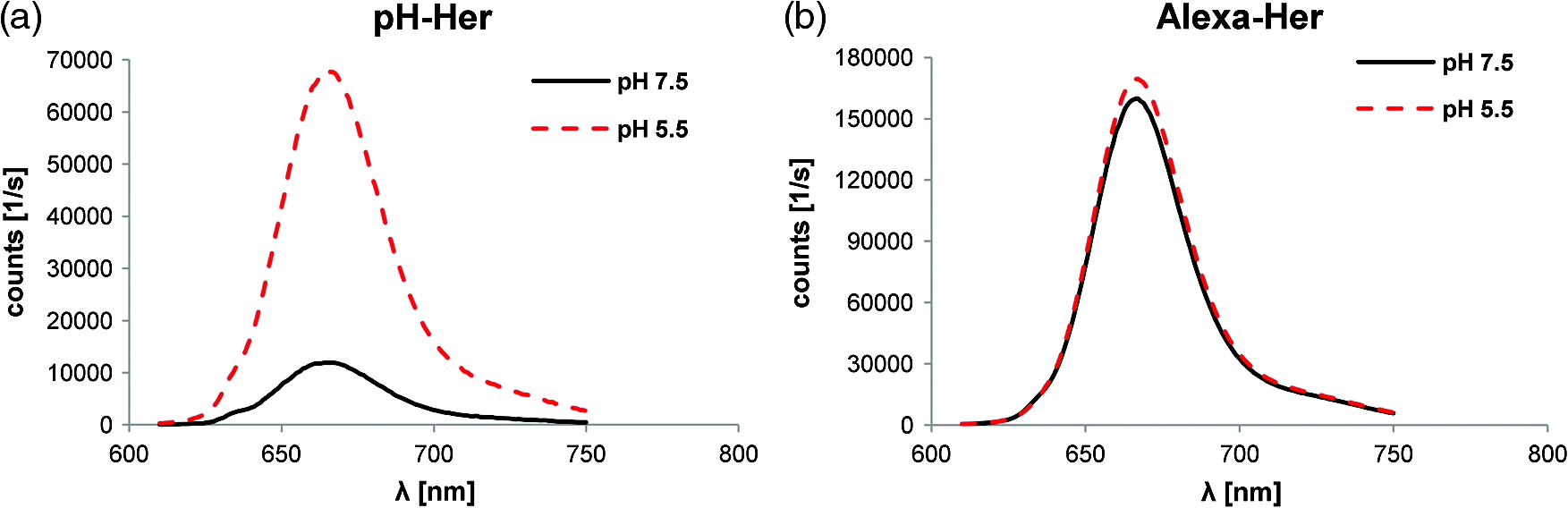 Fig. 3Fluorescence microscopy demonstrates internalization-dependent activation of pH-Her. Breast cancer cells grown on culture slides were incubated for 8 h with pH-Her or Alexa-Her. On the left panel, counterstain of cell nuclei with Hoechst 33342, in the middle, probe-derived signals, and on the right panel, merged images of the cell nuclei (blue) and the probe (red) are illustrated. (a), When incubated with KPL-4 cells at 37°C, pH-Her shows fluorescence only after receptor-mediated internalization (green arrow). (b), At 4°C, no signals from the pH-sensitive probe presumably bound to the cell membrane can be detected. (c), Alexa-Her shows fluorescence from the internalized probe (green arrow) and also membrane-derived fluorescence can be observed after 8 h of incubation at 37°C (c) and 4°C (d) (orange arrow, no internalization). Representative images of three independently performed experiments are presented. Bars represent 50 μm.  This article was corrected online on 6 August 2013. |


