|
|
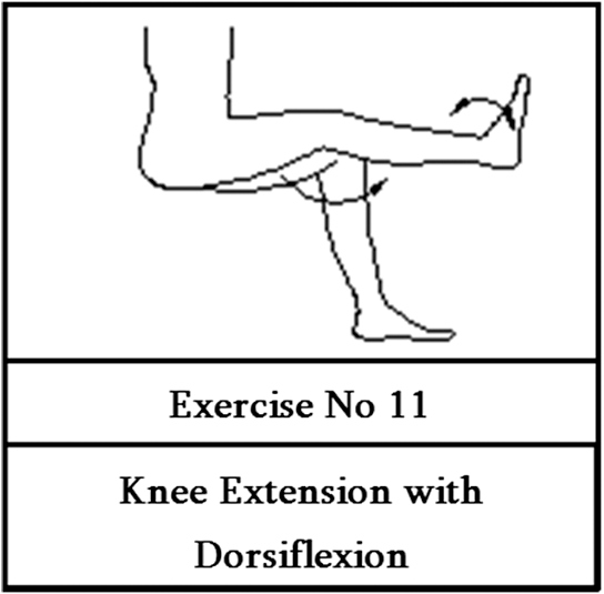
1.BackgroundImmobility of body or body parts due to surgery, pregnancy, illness, or long distance travel may subject one to the risk of deep vein thrombosis (DVT).1 DVT is a condition where the blood clot forms in the deep veins of the body, typically in the legs.2 The correlation between venous thromboembolism and air travel was established as early as 1954,3,4 and the occurrence of DVT in long distance air traveling passengers has increased since then.5 The magnitude of DVT risk in long distance air travel has been difficult to resolve due to the lack of adequate data. From observational studies, it is estimated that 1 out of 500 individuals above the age of 50 years who fly for 12 h can develop symptomatic DVT.6 A recent report has suggested that the risk increases by about 18% for each 2-h prolongation of travel duration.7 In legs, the arteries rely on the heart to force blood through them and the veins work against the gravity to transport blood with muscle contraction and relaxation to avoid DVT. Therefore, assisting blood circulation in the lower limb can reduce the risk of DVT especially during long hour flights. Several methods have been reported so far to increase venous circulation for DVT prophylaxis. Instrumental help has also been sought toward avoiding DVT, inducing small neuromuscular electrical impulses, which gently stimulate the common peroneal nerve behind the knee to activate the calf muscle pumps of the lower leg that return blood toward the heart.8,9 However, this method is better suited for elderly people struggling to walk or people with impaired locomotion. The easiest way to activate the calf muscle pump activity or to increase blood velocity in the veins is to manually exercise the calf muscles by performing certain exercises.10 Accordingly, most airline authorities recommend various leg exercises as a DVT preventative measure.11–13 Nonetheless, several studies conducted with the support of various sensor technologies suggest that not all exercises recommended by airline authorities induce active contraction of the calf muscle and therefore may not be beneficial to the passengers in prevention of DVT.14–17 In this context, the evaluation of the exercises suggested to avoid DVT using newer sensor methodologies is important. In the present work, fiber Bragg grating (FBG) sensors have been employed to evaluate the benefit of the set of airline exercises suggested to avoid DVT. This is a follow-up of the previously reported work on measuring surface strain on the calf muscle while performing exercises using FBG sensors.18 To validate the results obtained in the present study, the surface strain on the calf muscle, measured using FBG sensors during various exercises, has been compared against the blood velocity variation in the femoral vein of the leg obtained from a commercial color Doppler ultrasound (CDU) system. Various inherent advantages of FBG sensors—such as nonrequirement of energizing voltage, high special resolution, multiplexing capability, high strain resolution, sweat/humidity resistance, ability to be woven into a fabric to take the ergonomic shape of the body, etc.—make them suitable for biomechanical applications compared to other types of sensors, specifically in measurement of surface strain for evaluation of physical exercises. 2.FBG Sensors and InstrumentationFBG sensors have seen a considerable rise in the recent times, in their utilization in many varied applications.19–21 As mentioned earlier, the present work employs FBG sensors as the sensing element for dynamic measurement of strain on the surface of the calf muscle while performing the exercises suggested to avoid DVT. As the volunteered subjects might perform the exercises differently, an IMU which measures accelerations and rotational attributes has been used to confirm the accuracy/repetitiveness of exercises.22 Examining the blood velocity in the veins is a known technique for patients with chronic venous disease.23 Therefore, a commercially available CDU system is used on the femoral vein of the leg to measure change in blood velocity while performing exercises and the data obtained is compared against the results of the FBG sensors.24–26 3.Sensing MethodologyA 125-μm diameter, germania-doped, photosensitive, silica fiber has been used in the fabrication of FBG sensors of 3-mm gauge length, using the phase mask technique.20 The sensors are surface bonded on the medial belly of the right gastrocnemius muscle, using a skin friendly silicon-based adhesive as the bonding agent. The position and the placement of the FBG sensor have been optimized in consultation with the physician present during the test. The IMU is also strapped on the calf muscle using a Velcro strap. The probe of the CDU is placed against the femoral vein of the thigh to find the variation in the blood velocity during the performance of the exercises. The FBG sensor data is obtained in the form of change in the wavelength of reflected light from the FBG sensor, which is further converted into respective strain on the surface in which the sensor was mounted.27 In the present study, the experiments are conducted in a controlled environment in the laboratory. The atmospheric temperature remains fairly constant over the entire time of experimentation. The FBG sensor is attached to all the subjects at least 10 min before the actual data is recorded. This provides ample time for the FBG to attain thermal equilibrium with the body temperature of the subject, avoiding possible errors due to temperature drift. Data from all the three different sensing systems has been recorded simultaneously during the performance of the exercises by the subject. The data obtained from the IMU is used to find the accuracy of the repetition of the exercises performed by individual subjects with which an accuracy table is generated for all the exercises performed by each of the subjects. Each exercise is repeated for five times by all the subjects, and the corresponding strain data has been recorded using a FBG interrogation system from Micron Optics (SM 130–700), at a sampling rate of 1 kHz.28 The nature of strain generated (compression or tension) on the surface of the calf muscle depends on the movement pattern of the leg involved during each exercise. Irrespective of the nature of the strain generated on the calf muscle, whether it is tensile (positive strain) or compressive (negative), only magnitudes of strains are indicated for the purpose of illustration. 4.Experimental DetailsA detailed experimental procedure has been devised in the present work to illustrate the usefulness of FBG sensors for monitoring strain on the calf muscle. Figure 1 shows all the exercises considered for evaluation in the present study involving lower leg movements suggested by many of the airline authorities and biomechanical experts.11–13 4.1.SubjectsTwelve healthy subjects (six females and six males) have volunteered for the present work. They are aged between 24 and 28 years and are of varying body mass index (BMI mean: 25). Before starting the experiment, required ethical procedures have been followed. The subjects have been examined by a general medical practitioner one day prior to the experiment. All of them have been declared to be in good health condition to participate in the experiment. 4.2.ProcedureThe subjects are first briefed about the details of the experiment, such as the exercises to be performed, the posture in which it has to be performed, and the number of repetitions of each exercise. They are also instructed to repeat the exercises to the maximum extent of accuracy. To avoid fatigue error due to over straining the muscles of the subject, an interval of 5 min is given between two consecutive exercises. 4.3.Proposed ExerciseThe work by O'Donovan et al.14 has found that only few of the suggested exercises are beneficial in avoiding DVT. Since one of the authors associated with this work is an expert in yoga science, a new exercise has been proposed and evaluated. Figure 2 shows the proposed exercise in the present work, which involves extension of the knee with dorsiflexion of the ankle. The proposed exercise has also been evaluated for its performance in the same procedure adopted in the present work and the results were compared with the previously suggested exercises. 5.Results and DiscussionAs mentioned previously, data from all the three different sensing instruments has been recorded simultaneously during the performance of the exercise by the subjects. 5.1.IMU Data Processing for Accuracy Evaluation of ExercisesThe axes system of IMU (mounted on the calf muscle) has been shown in Fig. 3. All the measurements are made with respect to the IMU’s fixed axes system which outputs accelerations in , , axes and rotational attributes like pitch, yaw, and roll. As the IMU with a flat surface has been strapped on a curved calf muscle surface, initial offsets in the readings of the IMU are expected in the obtained data and normalized. IMU measurements of two exercises (exercise 1 and exercise 7) performed by a typical subject are presented for illustration in this section. Figure 4(a) shows the acceleration data along the three axes for five repetitions of the exercise, whereas Fig. 4(b) shows the angular rotational data acquired by the gyroscope in the IMU about the three axes. Similarly, accelerations and rotational attributes of exercise 7 are shown in Figs. 5(a) and 5(b), respectively. Fig. 4IMU data for exercise 1 with repetition of five trials for validation of accuracy. (a) Accelerations of exercise 1 and (b) rotational attributes of exercise 1. 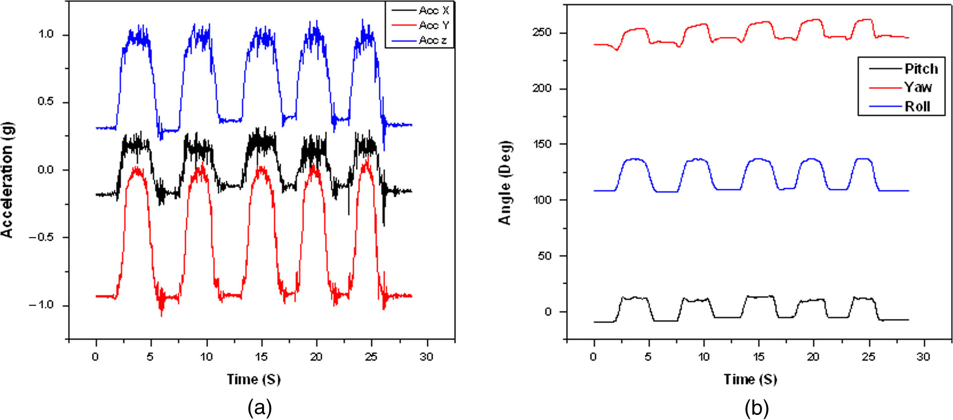 Fig. 5IMU data for exercise 7 with repetition of five trials for validation of accuracy. (a) Accelerations of exercise 7 and (b) rotational attributes of exercise 7.  From Figs. 4 and 5, it is evident that the subjects have performed the exercises quite accurately. Similar trends have been observed for all other exercises across all the subjects. The mean and standard deviation have been obtained for of all the six parameters (three axes and three rotational attributes). An average value of the standard deviation obtained for all the six parameters of each exercise is calculated and an accuracy table is generated, which is shown in Table 1. It can be seen from Table 1 that the accuracy levels obtained are in the acceptable range as the trials involved exercises from human beings. Table 1Accuracy table generated for all the exercises from inertial measurement unit (IMU) data.
5.2.FBG Sensor ResultsFigure 6 shows the real-time response of FBG sensor on the surface of the calf muscle for a typical exercise exerting tensile strain. Five distinct and sharp peaks corresponding to five repetitions of the exercise can be observed from the plot. Peak strain values recorded by all the five trials are comparable, signifying the consistency of the exercise performed by the subject. Fig. 6Real-time strain generated during an exercise recorded by the fiber Bragg grating (FBG) sensor on the calf muscle.  Figure 7 shows the box plot comparing each exercise for all subjects with mean, median, percentile ranges and spread of the strain on the calf muscle, with respect to time. From Fig. 7, it can be seen that among all the 11 exercises evaluated (including the newly suggested exercise), three distinctive groups of exercises evolve in terms of strains generated; group 1 comprises exercises 1 and 6, reading an average strain of that are found to be exerting more strain on the surface of the calf muscle compared to the rest of the suggested exercises. The second set (group 2) comprises exercises 4, 7, and 8, reading an average strain of on the surface of the calf muscle. Group 3 contains the rest of the exercises, namely exercises 2, 3, 5, 9, and 10 which exert comparatively minimal strain values in the range of on the surface of the calf muscle. Among all the evaluated exercises in this work, the newly suggested exercise, exercise 11 stands out best, exerting a mean strain of which is about 25% greater than the highest strain generated exercise (exercise 1) among the evaluated suggested airline exercises. 5.3.Study with CDU SystemFigure 8 shows CDU scans measuring the blood velocity in the femoral vein for a sample subject at a normal upright position and while performing typical exercises. Figure 8(a) shows the CDU scan illustrating blood velocity in the femoral vein when the subject is sitting in the normal upright position prior to performing any exercise. From Fig. 8(a), it can be seen that the velocity of blood is about . Figures 8(b)–8(d) show the blood velocity in the vein during exercises 1, 7, and 11, respectively, for two repetitions. It can be seen from Fig. 8 that the peak blood flow velocities for exercises 1, 7, and 11 are 126.99, 71.28, and , respectively. Similar scans have been obtained for all the other suggested exercises shown in Fig. 1. Fig. 8Color Doppler ultrasound (CDU) results showing velocity of blood: (a) blood velocity during normal leg position, (b) blood velocity during exercise 1, (c) blood velocity during exercise 7, and (d) blood velocity during exercise 11.  Figure 9 shows the plot between the blood velocity variations with respect to different exercises performed by the subject. It can be seen from this Fig. 9 that exercises 1 and 6 (group 1) are relatively more effective in terms of the variation in the blood velocity measured in the femoral vein. From the obtained CDU scans, exercises 4, 7, and 8 can be considered as group 2, which have shown intermediate magnitudes of variation in the blood velocity. On the same lines, the rest of the exercises, namely exercises 2, 3, 5, 9, and 10, comparatively show minimal variation in blood velocity as shown in Fig. 9. It is interesting to note that a similar grouping of exercises has also been obtained from the surface strain results of FBG sensor instrumentation (Fig. 7). A tabulation of the strain generated and the variation in blood velocity for all the exercises are shown in Table 2, which also depicts the groups evolved from both the FBG sensor and CDU techniques. Table 2Comparison of results from both fiber Bragg grating (FBG) sensor and color Doppler ultrasound (CDU) system for all the exercises.
Envelope comparison of Fig. 7 against Fig. 9 and from the exercise grouping obtained, confirming that the results of FBG sensors and the CDU system follow the same trend, match well with the work reported by O'Donovan et al.,14 confirming that the proposed FBG sensor methodology is reliable and accurate, thereby substantiating the potential of the FBG sensors in evaluating the suggested exercises. It can also be seen from Fig. 7 that the newly suggested exercise (exercise 11) generates more strain on the calf muscle and similarly more velocity in the femoral vein of the thigh (Fig. 9), proving its effectiveness in avoiding DVT compared to any of the previously suggested exercises; Exercise 11 proposed in this work records the strain and velocity which is 25% higher in terms of strain and blood velocity than exercise 1 which has recorded maximum among the suggested set of exercises (exercises 1 to 10). 6.ConclusionA new methodology using FBG sensors has been demonstrated to sense the strain on the surface of the calf muscle and to evaluate the benefit of the exercises suggested to avoid DVT during long distance flights. The results from the FBG sensors have been compared against the medically proven CDU system. In addition, an IMU has been used to measure the accuracy of repetition of each of the exercises performed by the volunteered subjects to ensure the reliability of the results obtained. Further, a new exercise has been proposed which is more effective as indicated by more strain on calf muscles and larger blood velocity in femoral vein. ReferencesP. Gispertet al.,
“Economy-class syndrome or immobile Traveler’s syndrome?,”
Archivos De Bronconeumologia, 42
(7), 373
–375
(2006). http://dx.doi.org/10.1016/S1579-2129(06)60546-6 Google Scholar
Deep Vein Thrombosis Anatomical Chart, Anatomical Chart Company, Chicago, IL
(2002). Google Scholar
J. Homans,
“Thrombosis of the leg veins due to prolonged sitting,”
N. Engl. J. Med., 250
(4), 148
–149
(1954). http://dx.doi.org/10.1056/NEJM195401282500404 NEJMAG 0028-4793 Google Scholar
I. S. SymingtonB. H. Stack,
“Pulmonary thromboembolism after travel,”
Br. J. Dis. Chest, 71 138
–140
(1977). http://dx.doi.org/10.1016/0007-0971(77)90097-3 BJDCAT 0007-0971 Google Scholar
K. J. Rothman,
“Thrombosis after travel,”
PLoS Med., 3
(8), 1206
–1207
(2006). http://dx.doi.org/10.1371/journal.pmed.0030300 PMLEAC 1549-1277 Google Scholar
S. C. Cannegieteret al.,
“Travel-related venous thrombosis: results from a large population-based control study (MEGA study),”
PLoS Med., 3
(8), 1258
–1265
(2006). http://dx.doi.org/10.1371/journal.pmed.0030307 PMLEAC 1549-1277 Google Scholar
D. Chandraet al.,
“Meta-analysis: travel and risk for venous thromboembolism,”
Ann. Intern. Med., 151
(3), 180
–191
(2009). http://dx.doi.org/10.7326/0003-4819-151-3-200908040-00129 AIMEAS 0003-4819 Google Scholar
R. E. Kaplanet al.,
“Electrical foot stimulation and implications for the prevention of venous thromboembolic disease,”
Thromb. Haemost., 88
(2), 200
–204
(2002). THHADQ 0340-6245 Google Scholar
P. D. Faghriet al.,
“Electrical stimulation-induced contraction to reduce blood stasis during arthroplasty,”
IEEE Trans. Rehabil. Eng., 5
(1), 62
–69
(1997). http://dx.doi.org/10.1109/86.559350 IEEREN 1063-6528 Google Scholar
K. Hitoset al.,
“Effect of leg exercises on popliteal venous blood flow during prolonged immobility of seated subjects: implications for prevention of travel-related deep vein thrombosis,”
J. Thromb. Haemost., 5
(9), 1890
–1895
(2007). http://dx.doi.org/10.1111/j.1538-7836.2007.02664.x JTHOA5 1538-7933 Google Scholar
(
(2013) http://www.jal.co.jp/en/health/flying/index.html#Prevention September ). 2013). Google Scholar
British Airways,
(2013) http://www.britishairways.com/en-in/information/special-assistance/medical-conditions September ). 2013). Google Scholar
Continental Airways,
(2013) http://www.united.com/web/en-US/content/travel/inflight/health.aspx September ). 2013). Google Scholar
K. J. Donovanet al.,
“Preliminary evaluation of recommended airline exercises for optimal calf muscle pump activity,”
Eur. J. Vasc. Endovasc. Surg. Extra, 12
(1), 1
–5
(2006). Google Scholar
K. J. Donovanet al.,
“The application of inertial and magnetic sensors to the monitoring of calf muscle pump activity,”
Med. Eng. Phys., 31
(1), 55
–60
(2009). http://dx.doi.org/10.1016/j.medengphy.2008.04.006 MEPHEO 1350-4533 Google Scholar
K. J. Donovanet al.,
“An inertial and magnetic sensor based technique for joint angle measurement,”
J. Biomech., 40
(12), 2604
–2611
(2007). http://dx.doi.org/10.1016/j.jbiomech.2006.12.010 JBMCB5 0021-9290 Google Scholar
K. J. Donovanet al.,
“Accelerometer based calf muscle pump activity monitoring,”
Med. Eng. Phys., 27
(8), 717
–722
(2005). http://dx.doi.org/10.1016/j.medengphy.2005.02.002 MEPHEO 1350-4533 Google Scholar
A. S. Guru Prasadet al.,
“A novel fiber bragg grating based sensing methodology for direct measurement of surface strain on body muscles during physical exercises,”
Int. J. Optomech., 6
(3), 189
–198
(2012). http://dx.doi.org/10.1080/15599612.2012.694982 1559-9612 Google Scholar
K. O. Hillet al.,
“Photosensitivity in optical fiber waveguides: application to reflection filter fabrication,”
Appl. Phys. Lett., 32
(10), 647
–649
(1978). http://dx.doi.org/10.1063/1.89881 APPLAB 0003-6951 Google Scholar
K. O. Hillet al.,
“Bragg gratings fabricated in monomode photosensitive optical fiber by UV exposure thorough a phase-mask,”
Appl. Phys. Lett., 62
(10), 1035
–1037
(1993). http://dx.doi.org/10.1063/1.108786 APPLAB 0003-6951 Google Scholar
W. W. Moreyet al.,
“Fiber Bragg grating sensors,”
Proc. SPIE, 1169 98
–107
(1989). http://dx.doi.org/10.1117/12.963022 PSISDG 0277-786X Google Scholar
(
(2013) http://files.microstrain.com/3DM-GX3-45-GPS-Aided-Inertial-Navigation-System-Data-Sheet.pdf September ). 2013). Google Scholar
C. M. Moloneyet al.,
“Haemodynamic study examining the response of venous blood flow to electrical stimulation of the gastrocnemius muscle in patients with chronic venous disease,”
Eur. J. Vasc. Endovasc. Surg., 31
(3), 300
–305
(2006). http://dx.doi.org/10.1016/j.ejvs.2005.08.003 1078-5884 Google Scholar
K. Yoshidaet al.,
“Color Doppler evaluation of valvular regurgitation in normal subjects,”
Circulation, 78
(4), 840
–847
(1988). http://dx.doi.org/10.1161/01.CIR.78.4.840 CIRCAZ 0009-7322 Google Scholar
W. D. FoleyS. J. Erickson,
“Color Doppler flow imaging,”
Am. J. Roentgenol., 156
(1), 3
–13
(1991). http://dx.doi.org/10.2214/ajr.156.1.1898567 AJROAM 0092-5381 Google Scholar
R. B. ChristopherM. D. Merritt,
“Doppler color flow imaging,”
J. Clin. Ultrasound, 15
(9), 591
–597
(1987). http://dx.doi.org/10.1002/(ISSN)1097-0096 JCULDD 0091-2751 Google Scholar
S. M. MelleR. M. Measures,
“A Bragg grating-tuned fibre laser strain sensor system,”
IEEE Photon. Technol. Lett., 5
(2), 263
–266
(1993). http://dx.doi.org/10.1109/68.196025 IPTLEL 1041-1135 Google Scholar
(
(2013) http://www.micronoptics.com/uploads/library/documents/Datasheets/Micron%20Optics%20sm130.pdf September ). 2013). Google Scholar
|