|
|
1.Introduction1.1.Energy-Based Surgical DevicesConventional suture ligation of blood vessels and tissue structures during open and laparoscopic surgery is a time-consuming and skill-intensive process. Recently, the alternative use of energy-based devices in place of sutures and mechanical clips (which leave foreign objects in the body) has enabled more rapid and efficient methods for vessel and tissue ligation. These energy-based devices can reduce surgical operative times1–3 and costs4–6 significantly. Several different types of energy-based instruments used in surgery today are capable of rapidly sealing blood vessels and tissue structures using thermal energy to denature proteins and reform the material by coagulation into seals that can withstand supraphysiological blood pressures. Clinically, most energy-based devices available for use are powered by ultrasonic or radiofrequency (RF) energy. Ultrasonic devices are indicated for coagulation and cutting of vessels up to 5 mm in diameter7,8 using thermal energy generated by rapid vibration of an acoustic waveguide. Alternatively, electrosurgical devices achieve hemostasis through the use of bipolar RF electrical current to generate heat resulting in thermal coagulation of vessels and are indicated for sealing vessels up to 7 mm in diameter.9–12 While some RF-based devices also use electrical and thermal energy to subsequently cut tissue, the majority of RF devices rely on a mechanical blade for this purpose. One of the main concerns with using energy-based devices in surgery, especially in procedures performed in tight spaces, such as prostatectomies and thyroidectomies, is the possibility of unintended thermal damage to adjacent critical tissue structures. All energy-based devices approved for clinical use produce varying amounts of thermal spread, which is defined as the amount of lateral thermal damage to tissue proximal to the seal site.13 Ultrasonic-based devices have been shown to produce less thermal spread than RF devices.14,15 However, the active jaw of ultrasonic devices can reach temperatures in excess of 200°C (Refs. 15 and 16) and take to cool to usable temperatures.17 Although RF devices are associated with larger thermal spread, their jaw temperatures are lower () than ultrasonic devices.15,16 These energy-based devices expedite normally labor-intensive surgical procedures, such as lobectomy,18 nephrectomy,3 gastric bypass,19 splenectomy,20 thyroidectomy,21 hysterectomy,22 and colectomy.23 However, both electrosurgical and ultrasonic devices have limitations, including the potential for undesirable charring and unnecessarily large collateral thermal damage zones (Table 1).14,24,25 Table 1Published ranges of mean values for seal time, burst strength, and thermal spread for commercially available vessel sealing devices (Refs. 14, 24, 26, 27, and 28).
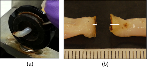
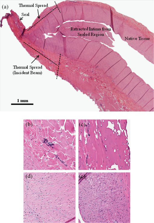
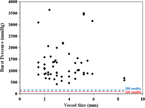
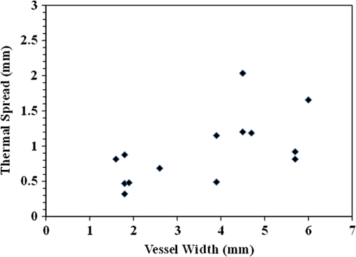
Note: RF, radiofrequency; US, ultrasonic. 1.2.Infrared Laser Vessel SealingIn other medical fields (e.g., ophthalmology and dermatology), lasers have been adopted as the surgical instrument of choice due to their enhanced level of precision compared to other energy-based devices. We hypothesize that a collimated infrared laser beam with an optical penetration depth closely matching that of the blood vessel diameter, and delivered over a short time period, may provide reduced lateral thermal spread in comparison to current energy sources. In a previous study, the efficacy of a number of infrared lasers with wavelengths of 808, 980, 1075, 1470, 1550, 1850 to 1880, and 1908 nm for sealing of blood vessels was reported.29 These preliminary studies demonstrated that the 1470 nm wavelength was capable of successfully sealing a wide range of porcine vessel sizes (1 to 6 mm) due, in part, to the laser’s intermediate optical penetration depth in soft tissues closely matching the thickness of the compressed vessels and its high power output. The purpose of this study was to investigate the high-power, 1470-nm infrared laser as an alternative energy source for rapid and precise sealing and cutting of blood vessels for potential use in open and laparoscopic surgical procedures where removal of diseased tissue is required. While our previous study demonstrated the feasibility of the 1470-nm laser wavelength with respect to the sealing of blood vessels, the specific objectives of this study were to demonstrate faster sealing for blood vessels with times comparable to current energy-based devices, incorporate transection of the sealed vessels, and demonstrate high vessel burst strengths to provide a safety margin for future clinical use. 2.Methods2.1.Tissue PreparationPorcine renal blood vessels were used for all of the laboratory studies. Fresh porcine kidney pairs were obtained (Animal Technologies, Tyler, Texas) and renal arteries were then dissected, cleaned of fat, and stored in physiological saline prior to same day use. Through careful dissection it was possible to surgically expose the entire vascular tree for each kidney, revealing numerous bifurcations, and multiple vessels with a wide range of diameters for testing. For this study, a total of 39 blood vessels were harvested ranging in outer diameter from 1.5 to 8.5 mm, and with a mean outer diameter of . Thirty of the vessels were sealed and cut resulting in 55 seal samples that were tested for burst pressures. Nine vessels were sealed and cut, providing 14 seal samples that were preserved for processing and histologic analysis. During the sealing process, the vessels were compressed to a fixed thickness of 0.4 mm. 2.2.Laser ParametersA 110-W diode laser with a center wavelength of 1470 nm (BrightLase Ultra-500, QPC Lasers, Sylmar, California) was used for all of the studies. Since water is the primary absorber of laser radiation in the near- to mid-IR spectrum, and soft tissues are composed primarily of water ( to 80% water content), the optical penetration depth (OPD) of 1470 nm radiation can be approximated by the water absorption coefficient ()30 using Beer’s Law (). This OPD also closely matches the compressed thickness of the blood vessels, which provides more efficient delivery of light energy to tissue. The laser was operated in long-pulsed mode with a pulse duration of 1 s, and at an output power of 110 W, providing 90 W of average power incident on the tissue surface for thermal coagulation and vaporization. Laser power output was measured after the beam shaping lenses using a power meter (EPM1000, Coherent, Santa Clara, California) and detector (PM-150, Coherent). 2.3.Experimental SetupThe bench top experimental setup was intended to mimic the tissue contact aspects of a surgical instrument while providing control over the experimental variables that may impact the future design of a handheld device. Some of the details of this experimental setup have been previously reported.29 Infrared laser radiation delivered through an armored, 400-μm-core, low-hydroxyl (OH), silica optical fiber patchcord (QPC Lasers) was collimated to a 10-mm-diameter beam using a 35-mm-focal-length (FL) plano-convex lens (LA1951-C, Thorlabs, Newton, New Jersey). A second, 100-mm-FL cylindrical lens (LJ1695RM-C, Thorlabs) then converted the circular spatial beam profile to a linear beam profile (Fig. 1). Fig. 1Diagram of linear beam-shaping optics, as well as respective blood vessel positions used for infrared laser sealing and cutting.  Two distinct spatial beam profiles for sealing and cutting were achieved by translating the vessel position 30 mm along the optical axis, thus moving the tissue out of and into the focus for sealing and cutting, respectively. This resulted in a change in the power density ratio (cutting/sealing) by a factor of 2.7. These two linear beams, aligned perpendicular to the vessel direction, are shown in Fig. 2 imaged with an IR spatial beam profiler (Pyrocam III, Spiricon, North Logan, Utah). The seal beam measured 9.5 mm long by 3.0 mm wide (power density of ) and the cut beam measured 9.6 mm long by 1.1 mm wide (FWHM) (power density of ). The vessel sample was sandwiched between a 1-mm-thick front glass slide and a 5-mm-wide back, metal faceplate with a glass slide insert, similar to our previously used bench top setup.29 The gap between the tissue contact surfaces when fully closed was fixed at 0.4 mm in the experimental setup to provide tissue compression closely matching the OPD of the laser wavelength. A force meter (25 LBF, Chatillon, Largo, Florida) then monitored the amount of force applied to the vessel sample, which was held constant at 36 N for this study. Laser energy was delivered in long-pulsed mode first with the seal beam dimensions for 1.0 s to create a thermal seal in the clamped vessel, and then the clamped vessel was translated 30 mm before cutting the vessel with an additional 1.0 s of laser irradiation at the beam focus. This large translation distance was a function of the cylindrical lens’s long focal length required for adequate optical element spacing in our bench top vessel compression setup. The sealing and cutting times of 1 s each were chosen based on earlier preliminary studies, which showed this to be the shortest period of time at maximum laser power that produced both strong seals and consistent cutting of a wide range of blood vessels. Fig. 2Dimensions of the sealing and cutting laser beam taken with an infrared beam profiler. The sealing beam measured (FWHM) and the cutting beam measured (FWHM), with a cutting/sealing beam intensity ratio of 2.7 (1080 versus ). The color scale representing beam intensity is in arbitrary units but is consistent in scale between the seal and cut beam measurements.  2.4.Burst Pressure MeasurementsVessel burst pressure measurements are considered a standard method for measuring vessel seal strength26,29,31 and was, therefore, used as the primary indicator of success for this study. The burst pressure setup consisted of a pressure meter (Model 717 100 G, Fluke, Everett, Washington), infusion pump (Cole Parmer, Vernon Hills, Illinois), and an iris clamp. The lumen of the vessel was placed over a cannula attached to the infusion pump. An iris was then closed to seal the vessel onto the cannula [Fig. 3(a)]. Deionized water was infused at a rate of and the pressure was measured with the pressure meter. The maximum pressure (in mmHg) achieved when the vessel seal bursts was then recorded. This process was repeated for both sides of cut vessels, providing two recorded burst pressures. A seal was considered successful if it exceeded both normal systolic blood pressure (120 mmHg) and malignant hypertension blood pressure (180 mmHg). The mean and standard deviation of the vessel burst pressures was then calculated from a total of 55 cut seal samples taken from 30 complete vessels harvested. Several vessel segments were too short to be properly mounted and tested in our burst pressure system. Fig. 3(a) Close-up view of sealed and cut 2.1 mm vessel enclosed in the iris during burst pressure testing. The vessel burst at a pressure of 882 mmHg. (b) Representative image of 5.2 mm vessel after sealing and cutting (white lines indicate extent of lateral thermal damage including the seal). This vessel burst at a pressure of 3447 mmHg. ().  2.5.Thermal Spread MeasurementsNine vessels ranging in diameter from 1.6 to 6 mm (mean of ) were sealed and cut, and of these vessels, 14 cut sides were salvaged for placement in formalin without bursting for at least 48 h before histological processing, and hematoxylin and eosin (H&E) staining. The histology samples were then imaged with an optical microscope (Olympus Model BX51) equipped with a digital camera (Olympus Model 71) for capture. Lateral thermal damage measurements were recorded using MicroSuite Biological Suite software system (Soft Imaging System GmbH). Thermal damage was measured from the proximal edge of the seal to the end of the coagulation zone where native tissue becomes apparent. For each individual cut seal sample, the thermal spread was averaged from both the front and back side of the vessel, representing a single data point. The mean and standard deviation of thermal damage was also calculated for all of the data points. Figure 4 shows a representative image of the H&E-stained longitudinal section of a cut vessel with the lateral thermal damage zones labeled. Note that the seal is not included in this measurement. Fig. 4(a) Representative H&E-stained histologic image of a 6-mm-diameter porcine renal blood vessel sealed and cut. The sealed region and the distance of lateral thermal spread from the seal are labeled. Thermal spread measured 1.83 mm on the top side and 2.17 mm on the bottom side. The elastic lumen is severed during lasing and retracts back into canal, leaving the stronger muscular layer to form the seal region. Histological markers (H&E staining, magnification) for healthy versus thermally damaged tissue are also shown: (b) Native adventitial collagen. (c) Denatured adventitial collagen. (d) Viable smooth muscle (media). (e) Thermally fixed smooth muscle (media).  3.ResultsFigure 3(b) shows a representative image of a blood vessel sealed and cut using the high-power 1470-nm laser. Burst pressure data points (each representing a seal side) as a function of the exact vessel diameter (mm) are plotted in Fig. 5. Normal systolic blood pressure (120 mmHg) and malignant hypertension blood pressure (180 mmHg) are labeled in the figure for reference. Mean burst pressures measured ( seal samples) with a minimum value of 415 mmHg recorded for a 3-mm-outer diameter (OD) vessel. As can be seen in the graph, there were no vessel sealing failures (). Fig. 5Vessel burst pressures plotted as a function of vessel size. Mean burst pressures measured ( seal samples) with a minimum value of 415 mmHg recorded for a 3-mm-outer diameter (OD) vessel. Normal systolic blood pressure (120 mmHg) and malignant hypertension (180 mmHg) thresholds are labeled for reference. All vessel seal samples tested burst above both of these thresholds.  Figure 6 shows the lateral thermal spread measured from the photographed histology samples ( seal samples, seal width not included in measurement) as a function of vessel size. Each individual data point represents the average thermal spread from the front and back side of the cut vessel. The mean for all of the data points shown in Fig. 6 was . 4.DiscussionThis study has shown that the high-power 1470-nm diode laser is capable of consistently producing strong seals in a wide range of porcine renal vessel sizes, ex vivo. A two-step technique combining both optical-based sealing and transection of vessels was demonstrated. The 2 s total irradiation time was comparable to other clinical RF and ultrasonic energy-based devices, which typically take 3 s or greater to seal and cut vessels (Table 1).26 It was postulated that larger seal regions would increase burst pressures. However, the beam widths used in this study were limited to 4 mm to allow for potential future integration into a standard, 5-mm-outer-diameter laparoscopic device. It should be noted that blood pressure through a vessel varies proportionally with the size of the vessel.32 In this study, all vessels tested exceeded both the normal systolic blood pressure of 120 mmHg and the severely elevated blood pressure levels of 180 mmHg experienced during malignant hypertension. Histologic measurements showed of thermal damage beyond the seal. These values are similar to or less than those typically created by using RF or ultrasonic devices (Table 1). Other energy devices typically produce mean burst pressures of to 930 mmHg with mean lateral thermal damage zones of 1 to 4 mm.14,24,25,26,27,28 However, for some of these devices, successful sealing of vessels have not been reported.21 Ultrasonic vessel sealing devices tend to produce less lateral thermal damage (1 to 1.9 mm), but require longer seal times (3.3 to 14 s), produce lower mean burst pressures (204 to 921 mmHg), and are limited to use on smaller vessels with diameter. RF devices require shorter treatment times (3.0 to 10 s), produce higher mean burst pressures (380 to 885 mmHg), and can be used on larger vessels up 7 mm diameter; however, they tend to produce greater lateral thermal damage (2.2 to 4 mm). The burst pressures and thermal damage zones measured after infrared laser sealing and cutting of vessels compares very favorably with those previously reported for both RF and ultrasonic devices (Table 1). In summary, this study describes several significant accomplishments. First, both sealing and cutting were consistently achieved in a wide range of vessels (1.5 to 8.5 mm) with a 100% () success rate. Second, the total laser irradiation time was only 2 s, comparable to other energy-based devices currently used in the clinic. Third, vessel burst strength averaged over 1300 mmHg, with the weakest vessel seal having a burst pressure of 415 mmHg, well above normal systolic blood pressure (120 mmHg). Although the bench top experimental setup described here was capable of simulating the tissue contact parameters of a surgical device, it is not suitable for in vivo animal surgical studies due to its large weight and size. Therefore, future work will require the design and testing of a miniaturized, handheld device suitable for testing in in vivo animal studies. 5.ConclusionsThis study demonstrates that a high-power 1470-nm diode laser is capable of rapid and precise sealing and cutting of a wide range of blood vessel sizes, ex vivo, with burst pressure values that are within the range of published values obtained from other commercially available energy-based instruments. Future work will focus on the development of a compact, handheld instrument for use during in vivo animal studies. AcknowledgmentsThis study was supported by a research grant from Covidien (Boulder, Colorado). Nicholas Giglio was supported in part by the Charlotte Research Scholars program at the University of North Carolina at Charlotte. The authors thank Christopher Cilip, Sarah Rosenbury, and Duane Kerr for their help with the initial stages of this project. Nicholas Giglio is now with the Biomedical Engineering Department at the University of Rochester. ReferencesZ. DingM. WableA. Rane,
“Use of Ligasure bipolar diathermy system in vaginal hysterectomy,”
J. Obstet. Gynaecol., 25
(1), 49
–51
(2005). http://dx.doi.org/10.1080/01443610400024609 JOGYDW 1364-6893 Google Scholar
B. LevyL. Emery,
“Randomized trial of suture versus electrosurgical bipolar vessel sealing in vaginal hysterectomy,”
Obstet. Gynecol., 102
(1), 147
–51
(2003). http://dx.doi.org/10.1016/S0029-7844(03)00405-8 OBGNAS 0029-7844 Google Scholar
C. Leonardoet al.,
“Laparoscopic nephrectomy using Ligasure system: preliminary experience,”
J. Endourol., 19
(8), 976
–978
(2005). http://dx.doi.org/10.1089/end.2005.19.976 JENDE3 0892-7790 Google Scholar
P. ManasiaA. AlcarazJ. Alcover,
“Ligasure versus sutures in bladder replacement with Montie ileal neobladder after radical cystectomy,”
Arch. Ital. Urol. Androl., 75
(4), 199
–201
(2003). Google Scholar
F. Romanoet al.,
“Laparoscopic splenectomy: ligasure versus EndoGIA: a comparative study,”
J. Laparoendosc. Adv. Surg. Tech. A., 17
(6), 763
–767
(2007). http://dx.doi.org/10.1089/lap.2007.0005 1092-6429 Google Scholar
P. W. Marcelloet al.,
“Vascular pedicle ligation techniques during laparoscopic colectomy. A prospective randomized trial,”
Surg. Endosc., 20
(2), 263
–269
(2006). http://dx.doi.org/10.1007/s00464-005-0258-7 SUREEX 1432-2218 Google Scholar
(
(2014) http://www.ethicon.com/healthcare-professionals/products/energy-devices/harmonic-ace-plus January ). 2014). Google Scholar
(
(2014) http://surgical.covidien.com/products/ultrasonic-dissection/sonicision January ). 2014). Google Scholar
(
(2014) http://www.ethicon.com/healthcare-professionals/products/energy-devices January ). 2014). Google Scholar
(
(2014) http://surgical.covidien.com/products/vessel-sealing#technology January ). 2014). Google Scholar
(
(2014) http://www.olympus-global.com/en/news/2012a/nr120321thunderbeate.jsp January ). 2014). Google Scholar
(
(2014) http://www.conmed.com/pdf%20files/altrus-brochure-mcm201072-revC.pdf January ). 2014). Google Scholar
R. H. LivingoodJ. A. VosJ. E. Coad,
“Hyperthermic tissue sealing devices: a proposed histopathologic protocol for standardizing the evaluation of thermally sealed vessels,”
Proc. SPIE, 7901 79010Y
(2011). http://dx.doi.org/10.1117/12.876861 PSISDG 0277-786X Google Scholar
G. W. Hrubyet al.,
“Evaluation of surgical energy devices for vessel sealing and peripheral energy spread in a porcine model,”
J. Urol., 178
(6), 2689
–2693
(2007). http://dx.doi.org/10.1016/j.juro.2007.07.121 JOURDD 0248-0018 Google Scholar
C. K. Phillipset al.,
“Tissue response to surgical energy devices,”
Urology, 71
(4), 744
–748
(2008). http://dx.doi.org/10.1016/j.urology.2007.11.035 0090-4295 Google Scholar
F. J. Kimet al.,
“Temperature safety profile of laparoscopic devices: harmonic ACE (ACE), Ligasure V (LV), and plasma trisector (PT),”
Surg. Endosc., 22
(6), 1464
–1469
(2008). http://dx.doi.org/10.1007/s00464-007-9650-9 SUREEX 1432-2218 Google Scholar
H. R. Govekaret al.,
“Residual heat of laparoscopic energy devices: how long must the surgeon wait to touch additional tissue?,”
Surg. Endosc., 25
(11), 3499
–3502
(2011). http://dx.doi.org/10.1007/s00464-011-1742-x SUREEX 1432-2218 Google Scholar
M. Garanciniet al.,
“Bipolar vessel sealing system vs clamp crushing technique for liver parenchyma transaction,”
Hepatogastroenterology, 58
(105), 127
–132
(2011). Google Scholar
A. StoffM. A. ReichenbergerD. F. Richter,
“Comparing the ultrasonically activated scalpel (harmonic) with high-frequency electrocautery for postoperative serious drainage in massive weight loss surgery,”
Plast. Reconstr. Surg., 120
(4), 1092
–1093
(2007). http://dx.doi.org/10.1097/01.prs.0000278224.14221.c9 PRSUAS 0032-1052 Google Scholar
Y. Kurtet al.,
“New energy-based devices in laparoscopic splenectomy: comparison of Ligasure alone versus Ligasure and Ultracision together,”
Surg. Pract., 16
(1), 28
–32
(2012). http://dx.doi.org/10.1111/j.1744-1633.2011.00577.x 1744-1625 Google Scholar
Y. Ponset al.,
“Comparison of LigaSure vessel sealing system, harmonic scalpel, and conventional hemostasis in total thyroidectomy,”
Otolaryngol. Head Neck Surg., 141
(4), 496
–501
(2009). http://dx.doi.org/10.1016/j.otohns.2009.06.745 0194-5998 Google Scholar
J. KroftA. Selk,
“Energy-based vessel sealing in vaginal hysterectomy: a systematic review and meta-analysis,”
Obstet. Gynecol., 118
(5), 1127
–1136
(2011). http://dx.doi.org/10.1097/AOG.0b013e3182324306 OBGNAS 0029-7844 Google Scholar
M. Adaminaet al.,
“Randomized clinical trial comparing the cost and effectiveness of bipolar vessel sealers versus clips and vascular staplers for laparoscopic colorectal resection,”
Br. J. Surg., 98
(12), 1703
–1712
(2011). http://dx.doi.org/10.1002/bjs.v98.12 BJSUAM 0007-1323 Google Scholar
G. R. Lambertonet al.,
“Prospective comparison of four laparoscopic vessel ligation devices,”
J. Endourol., 22
(10), 2307
–2312
(2008). http://dx.doi.org/10.1089/end.2008.9715 JENDE3 0892-7790 Google Scholar
P. A. Suttonet al.,
“Comparison of lateral thermal spread using monopolar and bipolar diathermy, the Harmonic scalpel, and the Ligasure,”
Br. J. Surg., 97
(3), 428
–433
(2010). http://dx.doi.org/10.1002/bjs.v97:3 BJSUAM 0007-1323 Google Scholar
W. L. Newcombet al.,
“Comparison of blood vessel sealing among new electrosurgical and ultrasonic devices,”
Surg. Endosc., 23 90
–96
(2009). http://dx.doi.org/10.1007/s00464-008-9932-x SUREEX 1432-2218 Google Scholar
J. Landmanet al.,
“Evaluation of a vessel sealing system, bipolar electrosurgery, harmonic scalpel, titanium clips, endoscopic gastrointestinal anastomosis vascular staples and sutures for arterial and venous ligation in a porcine model,”
J. Urol., 169
(2), 697
–700
(2003). http://dx.doi.org/10.1016/S0022-5347(05)63995-X JOURDD 0248-0018 Google Scholar
B. Personet al.,
“Comparison of four energy-based vascular sealing and cutting instruments: a porcine model,”
Surg. Endosc., 22 534
–538
(2008). http://dx.doi.org/10.1007/s00464-007-9619-8 SUREEX 1432-2218 Google Scholar
C. M. Cilipet al.,
“Infrared laser thermal fusion of blood vessels: preliminary ex vivo tissue studies,”
J. Biomed. Opt., 18
(5), 058001
(2013). http://dx.doi.org/10.1117/1.JBO.18.5.058001 JBOPFO 1083-3668 Google Scholar
G. M. HaleM. R. Querry,
“Optical constants of water in the 200 nm to 200 μm wavelength region,”
Appl. Opt., 12 555
–563
(1973). http://dx.doi.org/10.1364/AO.12.000555 APOPAI 0003-6935 Google Scholar
W. W. Hopeet al.,
“An evaluation of electrosurgical vessel-sealing devices in biliary tract surgery in a porcine model,”
HPB, 12
(10), 703
–708
(2010). http://dx.doi.org/10.1111/hpb.2010.12.issue-10 HPBSE9 0894-8569 Google Scholar
I. W. ShermanV. G. Sherman, Biology: A Human Approach, Oxford UP, New York
(1979). Google Scholar
|


