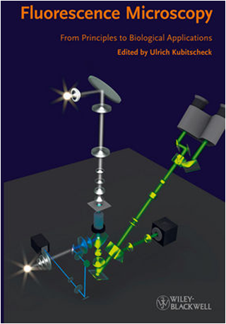
Ulrich Kubitscheck, Ed., 539 pages, ISBN: 978-3-527-32922-9, Wiley-Blackwell, Weinheim, (2013), $169.95, hardcover.
Reviewed by Barry R. Masters, Fellow of AAAS, OSA, and SPIE.
Microscopes are tools invented by humans to study specimens at spatial and temporal resolution that exceed the human eye. Today the researcher has a wide range of microscopes to employ in order to answer carefully poised experimental questions. The key is to match the research questions with the appropriate microscope. In order to optimize the success it is necessary to understand the physical principles of the microscope, how to properly align the instrument, and how to correctly use the instrument. The object of the investigation or measurement is also a major consideration as well as the preparation of the specimen, the interaction of the specimen, and the radiation from the microscope. Often the words “quantitative microscopy” appear in publications; but all measurements contain errors, and the critical investigator must be able to understand the sources of error in a measurement and, if possible, mitigate the magnitude of the errors. Validation of microscopic measurements across disparate types of microscopes is a useful approach. The critical elements in optical microscopy include: optical aberrations, sampling errors, nonphysiological specimen preparation, such as over expression of genetic fluorescent proteins, the deleterious effects of the light source on biological specimens, nonlinear effects in the detector and amplifiers, and artifacts of image analysis and image interpretation.
From the beginning, microscopes were plagued by artifacts and problems of contrast and resolution. Methods to enhance the contrast of the specimen have a long history. The use of fluorescent dyes to enhance contrast in bacteria resulted in Robert Koch receiving the 1905 Nobel Prize in Medicine. The development of fluorescent probes has resulted in a plethora of advances in biology and medicine, but they are not without their disadvantages; i.e. photodamage, photobleaching, and other nonphysiological effects on the specimens. Modern advances in live-cell imaging and deep-tissue imaging, and in vivo imaging of cells, tissues, and organs, such as the living brain and organisms, are furthering our advances in biomedicine. We should not forget that nonfluorescent methods to enhance contrast provide important alternative approaches. One key example is the use of Golgi staining of specific neurons in the central and the peripheral nervous system; these stains sparsely affect specific neurons in the nervous system and the contrast is derived from changes in the optical properties of the stained neurons. Our understanding of the morphology, development, and regeneration of the nervous system derived form the investigations of Santiago Ramón y Cajal, who used and improved the silver staining technique of Camillo Golgi; they both shared the 1906 Nobel Prize for Physiology or Medicine. Other important examples of optical contrast enhancement are the invention of phase contrast and differential interference contrast microscopes. While fluorescent and nonfluorescent probes are commonly used in modern microscopy, there are new “probeless” microscopes that form the contrast without the need for and the artifacts of fluorescent probes.
The genesis of Fluorescence Microscopy: From Principles to Biological Applications was the 2009 microscopy workshop held at Rockefeller University. The editor contacted the speakers of the workshop and they contributed the book’s chapters. A significant omission is the lack of a chapter on multiphoton microscopy; the term is mentioned several times in the book without a listing in the index, but there is no discussion of its physical basis, its unique advantages for deep imaging in vivo, and its numerous biological applications, as evidenced by the number of publications and citations related to this microscopic technique.
The anticipated audience for the book is “students and researchers with little background in physics.” This statement is too vague to convey the level of mathematics and physics that the reader requires to comprehend the text. I think the book is suitable for undergraduate and graduate students who work and study in the biomedical fields and wish to understand the principles of modern optical microscopy, the instruments, and their limitations. The contributors have isolated the physics related to each chapter into blue boxes that the reader can chose to read depending on his or her individual background in physics. In this manner, chapters read as self-contained units even without the detailed information in the blue boxes.
This book is excellent as a pedagogical textbook and that separates it from a large number of similar books. This quality is achieved by several features; the most important is the clarity of the writing and the many figures. Many of the figures are produced in color and that helps the reader’s comprehension. The references that accompany each chapter serve to augment the text, but they date from 3 years; thus, more recent references and review articles are missing. Some contributors have marked key references with a gray box to highlight their significance.
The text is supplemented with a useful appendix that serves as a practical guide to optical alignment; this is key to the optimal function of microscopes. Lasers are often used as light sources for many microscopic techniques; therefore, an appendix on laser safety is an important omission. Another chapter on the physics of lasers would benefit the reader if it was included. In addition to an index, which omits several key terms, i.e. Kasha’s Rule, there is a website for the book that contains all of the figures (which can be used for lectures) and six movies, each of about ten-second duration. The movies add little to the book. Several key terms are inadequately described. For example, the term adaptive optics is covered in one sentence. The inclusion of current review articles in the references would compensate for the brevity of the discussion. The chapters differ in the quality and the number of references; some chapters have an extensive selection of articles and reviews together with in-text citations; other chapters have a paucity of references.
The unique pedagogical value of Fluorescence Microscopy is the clear writing, the explanations of the physical concepts, the clear figures, and the discussion of problems and limitations of each microscopic technique. The editor and the contributors have produced a highly recommended textbook that is very readable with a good balance of theory, instrumentation, and applications.


