|
|
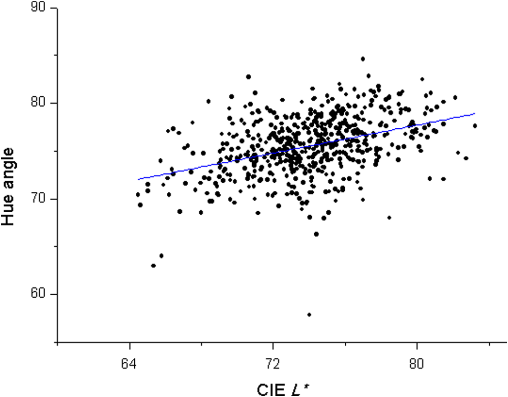
1.IntroductionColor is a subjective sensation and as such is difficult to use in a quantitative study.1 However, varied color measuring instruments such as spectrophotometer, colorimeter, spectroradiometer, and image analysis of digital images were used to quantitatively express the color of teeth and orofacial tissues.2,3 Color can be described according to the Munsell color specifying system in terms of Hue, Value, and Chroma.4 Human teeth occupy a range of Munsell color space approximately Hue from 8 yellow-red to 3 yellow, Value from 6/ to 8.5/ and Chroma from /1 to /5.5 Today, the most widely used quantitative color specification system is the Commision Internationale de l’Eclairage (CIE) “Lab” system.6 In this system, the CIE stand for lightness (achromatic coordinate), green-red coordinate (negative is green and positive is red), and blue-yellow coordinate (negative is blue and positive is yellow), respectively. Teeth color can also be described within the cylindrical color space.7 is the lightness that is defined as the perceived brightness, ranging from black to white, and is quantified on a scale from 0 to 100. is the chroma that describes the saturation of a surface color or the degree of visual difference from neutral grey. Hue angle () forms a continuous circular scale and is indicated in angles from 0 to 360 deg. The cylindrical color space can be transformed into the CIE Lab color space and vice versa.7 Tooth color is decided by a combination of the intrinsic color resulting from the interaction of light with the tooth structure and the presence of extrinsic stains.8,9 Light scattering and absorption within enamel and dentine give rise to the intrinsic color of a tooth. Since enamel is relatively translucent, the optical properties of dentine play a major role in determining the overall tooth hue and chroma.3,10 Optical properties of human teeth are also influenced by their external configuration. As to the regulation of tooth shape, epithelial-mesenchymal interactions establish the morphology of the dentine surface upon which enamel will be deposited. Starting with the onset of amelogenesis beneath the future cusp tips, the shape of the enamel layer covering the crown is determined by several growth parameters.11 The dimension, shape, and surface structure of a tooth generate light reflection patterns which influence the overall tooth color. Knowing that the amounts of reflected and absorbed light depend on the thickness and translucency of these tissues, it is evident that the thicknesses of enamel and dentine affect tooth color.12 Tooth development is determined by genetic and environmental factors.13 Developmental enamel malformations can be caused by genetic or environmental factors. A systemic condition during tooth development, such as high fever, can produce a pattern of enamel defects in dentition.14 Inherited diseases may influence the thickness of enamel or the mineral and organic content of the enamel; therefore, they can affect tooth color.15 Dental fluorosis processes are likely to be involved in the genetic determination of enamel thickness and conceivably may influence tooth color.16 The process of enamel maturation continues following tooth eruption, so that the erupted tooth can change color over time.15 Adult teeth also record environmental and traumatic events, as well as the effects of disease and ageing.13 Human teeth are sensitive to the influences experienced during adult life, particularly those nutritional, metabolic, traumatic events and diseases that lead to changes in coloration.8,17 Tooth enamel is composed of individual crystallites that are larger and more oriented than other mineralized tissues. Mature enamel crystallites are composed of non-stoichiometric carbonated calcium hydroxyapatite.14 Tooth color is regulated by the size of its enamel hydroxyapatite crystals.9 However, the exact genetic and/or environmental mechanisms that decide the tooth color are not thoroughly understood at this time. Human tooth color has been measured with varied measuring methods.3,7,18,19 In general, these studies showed a large range in the CIE , and values, but consistently showed that there was a significant contribution of the CIE value or yellowness to tooth color.3 The mean values for the CIE , , and of teeth were reported as 70.7, 4.3, and 17.5, respectively, for adults and teenagers.19 Tooth color is also a rich source of information concerning health and genetic quality.13 Age and gender were the most important factors associated with human tooth color, and about half of the investigated population suffered from some tooth discoloration and were dissatisfied with their tooth color.19 Based on a study on the demographic, behavioral, and treatment parameters that impacted tooth color and its perception, although age contributed to objectively measured tooth discoloration, personal satisfaction with tooth color was age-independent.20 Dental appearance plays a key role in the way we develop a first impression of another person. A perceptible change in the lightness of teeth was the strongest factor associated with the dental attractiveness stereotype.21 On the other hand, it was reported that the presence of yellowed coloration had negative effects on ratings of attractiveness based on an experiment on the effects of digital manipulations of teeth color.13 This result indicated that these color-induced alterations in ratings of attractiveness were mediated by increased/decreased yellowing rather than whitening per se. There have been studies that determined the correlations between the tooth color and age/gender,22,23 color of skin, hair and eyes,24,25 and facial features and gender.26 Several of these results were applied in the prediction of the teeth color from the correlated values in the studies. Color measurements of anterior teeth showed that a tooth did not have a single uniform color and the middle site appeared to represent the tooth color best.27 Although it was reported that the Munsell Hue, Value, and Chroma of human teeth were not independent of one another and were highly correlated with each other,5 the Munsell color attributes are basically not quantitative values and do not have physical meanings such as the threshold values for visual or perceptual interpretation. Since the correlations based on the Munsell notations were not quantitative, this result could not have quantitatively provided the degree of correlation or harmonization of tooth color. In the present study, it was assumed that there should be significant correlations among the quantitative color coordinates of each tooth because the color of each tooth is influenced by the genetic, congenital, metabolic, chemical, infectious, and environmental factors of each person.17 The working hypothesis assumed in the present study was that the three numerical color coordinates, such as the CIE , and coordinates and the lightness (CIE ), chroma () and hue angle () coordinates, would show significant correlations. Therefore, the influences of the color coordinates on the other coordinates were determined using the mean values of the same type of tooth in the same person as the raw data. The purpose of this study was to determine whether there were significant correlations between the numerical color coordinates of the human teeth. 2.Material and Methods2.1.Tooth Color MeasurementThe tooth color of 47 volunteers, older than 19 years old, was measured (). Approval was obtained from the institutional review board and informed consent was obtained from each of the volunteers. Through clinical examination of oral health conditions of each volunteer, it was confirmed that they did not have caries, abraded lesions, or restorations in any of the six maxillary and six mandibular anterior teeth. Their periodontal conditions were in the range of fair to good. Eight males and 39 females were included, and their mean age was 29.5 () and 29.0 (), respectively.18 The color of 12 maxillary and mandibular anterior teeth was measured by Shade Vision System (X-rite, Grandville, MO, USA). This system is a commercially available shade taking device that provides an accurate colored “contour map” image of the tooth. It is essentially a colorimeter that utilizes image-grabbing technology. It comprises a handheld measuring device that is used to scan the tooth surface together with a docking station linked to a computer and associated software.28 This device is calibrated with a reflection standard and a searchlight illuminator is used as the light source.29 This device showed excellent repeatability.29,30 To exclude the influence of environmental light, all the measurements were performed between 5 PM to 6 PM at a dental unit chair not receiving any direct sunlight, and one dentist measured the color using the same measurement protocol. The aperture head was contacted at the center of each tooth. Measurements were repeated three times and averaged. 2.2.Color Coordinates of TeethTwo color coordinate systems such as the CIE , and system and the lightness (CIE ), chroma (), and hue angle () system were used. Chroma was calculated as , and hue angle was calculated as .6 2.3.Statistical AnalysisCorrelations between each pair of the three color coordinates in each system and other correlated pairs from the CIE , , and chroma were determined with a linear regression analysis (). In these regressions, pairwise comparisons of the color coordinates based on the same tooth in the same person were made. Pearson correlation coefficient and regression equation were calculated for each pair. Within each of the two color coordinate systems, the predictability of one color coordinate from the values of two other coordinates was estimated with a multiple regression analysis (). For example, in the CIE , and system, the CIE coordinate was set as a dependent variable and the CIE and coordinates were set as independent variables. These regressions were performed sequentially by switching the variables. To eliminate the impact of interrelated independent variables, the variable which showed the lower standardized partial correlation coefficient () was not included in the regression equation when the tolerance between two influencing variables was lower than 0.30.31 3.ResultsThe mean and distribution ranges of human teeth color coordinates were as follows; CIE and , and 5.0 (1.5), and 19.4 (4.0), chroma and 20.1 (4.2), and hue angle and 75.5 deg (3.1). Tooth color was in the first quadrant of the CIE and plane because the CIE and values were positive; therefore, the hue angles were also in the range of 57.8 to 84.6 deg. Correlations between the CIE , and or coordinates of teeth are presented in Fig. 1. Except for several isolated points, they showed linear regression trends. Correlation between the CIE and values is presented in Fig. 2, which showed a high positive correlation (correlation coefficient; ). The correlation between the lightness (CIE ) and chroma (Fig. 3) was negative, and the correlation between the lightness and hue angle (Fig. 3) was also observed. Fig. 1Correlation between Commission Internationale de l’Eclairage (CIE) coordinates of human teeth.  Correlation coefficients and regression equations between each pair of the CIE , and coordinates, and the lightness, chroma, and hue angle coordinates are presented in Table 1. When the CIE was set as a dependent variable (set as ), the correlation coefficients were in the range of to 0.40. The CIE was negatively correlated with the CIE , , and chroma but positively with the hue angle. When the CIE was set as a dependent variable, it was positively correlated with the CIE and chroma. When the CIE was set as a dependent variable, it was positively correlated with the chroma (). In the lightness, chroma and hue angle system, the lightness showed a negative correlation with the chroma and a positive correlation with the hue angle. The chroma and hue angle showed no significant correlations. Table 1Correlations between each pair of CIE L*, a*, and b* coordinates and lightness (CIE L*), chroma (C*ab), and hue angle (h°) coordinates.
Multiple regression results based on all the data are presented in Table 2. The coefficient of determination () means the square of the multiple correlation coefficient and indicates how well the data points fit a statistical model. In the CIE , and system, the coefficient of determination was in the range of 0.41 to 0.57; and in the lightness, chroma, and hue angle system, this value was in the range of 0.23 to 0.43. In this table, means the standardized partial correlation coefficient for each included variable with the dependent variable when statistically significantly correlated independent variables (predictors) are included. The magnitudes of coefficients of determination for the CIE , and system were higher than those of the lightness, chroma, and hue angle system. Table 2Multiple regression results among CIE L*, a* and b*, and lightness (CIE L*), chroma (C*ab), and hue angle (h°) based on all data.
Differences in the multiple regression results for the maxillary or mandibular anterior teeth are presented in Table 3. The coefficients of determination for the maxillary teeth were in the range of 0.22 to 0.62 and those for the mandibular teeth were 0.23 to 0.50. The orders of included variables changed in several cases. Table 3Differences in the multiple regression results in maxillary and mandibular teeth.
4.DiscussionThe working hypothesis that the three numerical color coordinates of human teeth would show significant correlations was partially accepted because, in both of the color coordinate systems, the three color coordinates showed significant correlations except for the chroma and hue angle pair. Because the chroma refers to the intensity of the hue and the hue angle refers to the CIE and ratio, they indicate different aspects of the chromatic coordinates in the same plane; therefore, they did not show significant correlation. As for the CIE and distribution ranges of teeth in the CIE and plane, these coordinates were in the first quadrant, which indicates that tooth hue is basically in the red and yellow region not far from the neutral gray axes. As presented in Figs. 1 to 4, the color coordinate pairs showed linear regression trends except for several isolated points. As for the isolated points, exceptional or abnormal color coordinates of particular teeth caused by genetic and/or other environmental factors, measurement error, undetected discoloration, or staining might have resulted in these deviated points. As indicated in Figs. 1 and 3, a lighter tooth (higher CIE value) showed lower (in this case, close to neutral grey) and (also close to neutral grey) values and accordingly lower chroma values. When the CIE was set as a dependent variable, the correlation coefficients were in the range of to 0.40, and it was negatively correlated with the CIE , , and chroma, but positively with the hue angle (Table 1). It is not known whether the lightness of teeth directly influenced the chromatic color coordinates and vice versa. Further analytical study for the theoretical and biological backgrounds of these phenomena should be performed. The correlation coefficient between the CIE and chroma was very high (). Therefore, the chroma was mainly determined by the CIE , and collaterally by the CIE (). Multiple regression results based on all the data are presented in Table 2, and the differences in the multiple regression results between the maxillary and mandibular teeth are presented in Table 3. The coefficients of determination for the maxillary teeth were generally higher than those for the mandibular teeth. Therefore, the predictability of a missing color coordinate calculated with the other two coordinates in the maxillary teeth would be higher than that in the mandibular teeth. Also, compared with the results based on all the data (Table 2), the predictability of a missing coordinate in the maxillary teeth was higher than all the teeth included condition except for hue angle. As for the differences by gender, since the number of male volunteers was limited, it seemed unreasonable to directly compare the values. However, the trends were similar in both male and female groups. The coefficients of determination for males were generally higher than those for females, which might reflect the limited numbers of isolated points in the male group because a small number of teeth was included (12 teeth for each of eight male volunteers = 96 teeth). The correlation coefficient between the CIE and was 0.66 in the present study (). Correlations between the color coordinates of human teeth were already mentioned in previous studies from different viewpoints.2,5,32 In a study on the correlations of the Munsell color attributes in natural teeth, it was reported that three attributes were closely correlated.5 Although the correlation coefficient between Hue and Value was reported as 0.80, direct comparison of the results with those of the present study seemed meaningless because the parameters used in this previous study were not truly quantitative. In another study, the validity of a non-contacting spectroradiometer for the color determination of craniofacial structures was assessed.2 Although the authors did not perform the correlation analyses for their color coordinate data of three anterior teeth, it was presented in the graphs that the CIE and values showed positive correlations, the CIE and values showed negative correlations and the CIE and values showed weak negative correlations. These results were similar to those of the present study. Therefore, statistical analyses for these coordinates were performed in the present study. As to the correlation between the CIE and , there was a report based on a theoretical approach.32 In the CIE color space, the chromaticity coordinates are judged separately from the lightness parameter, the latter being represented by an additional axis.6 This means that the uniform CIE Lab color system implies strong coherences between the chromaticity data in terms of the CIE and as both are located on the same plane and should, therefore, display a strong dependency. In the present study, in addition to the correlation between the CIE and values, correlations between the CIE , chroma and hue angle were also confirmed. Heritability estimates, compared with environmental effects of treatment responses to the teeth bleaching in twins were determined.33 Whitening treatment responses were highly heritable for and , but not for , which was essentially modulated by environmental factors. From this study, it might be postulated that the CIE and , and accordingly chroma, are highly correlated and the CIE value is a separate parameter. As previously mentioned, the theoretical and biological backgrounds for these phenomena should be further studied. The influence of variations in enamel ultrastructure on the optical properties of teeth was investigated.9 Tooth shade, enamel chemical composition, and crystallography were assessed. Pearson correlation analysis demonstrated that tooth hue was associated with enamel hydroxyapatite (HA) crystal size (), tooth chroma was associated with enamel HA carbon content (), and tooth lightness was associated with both enamel HA crystal size () and the degree of HA carbon content (). These findings are of great relevance in dentistry since it provides a better understanding of tooth esthetics. Although this study partially provided biological mechanisms of tooth color determination, the correlations among the color parameters were not discussed in this paper. As for the theoretical approaches for tooth color determination, measured reflectance spectra of natural enamel and dentine specimens were in good agreement with the Kubelka–Munk theoretical values.34 The reflectance spectra of natural enamel and dentine sections were measured and this allowed the calculation of the Kubelka–Munk scattering and absorption parameters, which were compared with those of the restorative materials.35 Although biological mechanisms for tooth color determination should be further studied, there have been several studies on the influence of filler contents and other factors on the shade of dental resin composites. These results might explain some part of the biological or theoretical tooth color determination mechanisms. It was reported that the lightness of resin composites was highly correlated with the amount of filler, scattering coefficient, and refractive index (). But the correlation coefficients between the amount of filler and chroma/hue were moderate ().36 Since the scattering and absorption characteristics influence the color of resin composites, the size and volume fraction of fillers should be controlled for the best color reproduction, considering the refractive indices of the filler and resin matrix.37 The limitations of the present study were (1) although the number of investigated teeth was 564, a larger number might have provided more trustworthy results and (2) distributions of gender and age of the volunteers were limited. Practically, the results of the present study can be applied to the shade guide design, prediction of missing color coordinate of teeth, analyzing the mechanisms of tooth development/shade determination and acquired disease reflected in tooth color and the tooth whitening effect evaluation. 5.ConclusionsBased on the results of the present study, all the color coordinate pairs of human vital teeth showed significant correlations except for the chroma and hue angle pair. Lighter teeth (higher value) showed lower (closer to neutral grey) and (also closer to neutral grey) values and accordingly lower chroma. The CIE was positively correlated with the CIE and chroma. Therefore, it was postulated that three color coordinates of a tooth are harmonized within certain color attribute ranges and a lack of correlations in these coordinates might indicate external/internal discolorations and/or the anomaly of a tooth. Deviations away from normal harmonized range of tooth color coordinates might have negative effects on self-satisfaction and the social attractiveness of a human. AcknowledgmentsThis study received the approval of the IRB (Internal Review Board) of Seoul National University Dental Hospital, Seoul, Republic of Korea (CME05002). Author declared no conflict of interest. ReferencesN. DevosG. WillemsR. Wood,
“Objective human tooth color measurements as a means of determining chronologic age in vivo and ex vivo,”
J. Forensic Odontostomatol., 27
(2), 2
–8
(2009). Google Scholar
D. J. Gozalo-Diazet al.,
“Measurement of color for craniofacial structures using a 45/0-degree optical configuration,”
J. Prosthet. Dent., 97
(1), 45
–53
(2007). http://dx.doi.org/10.1016/j.prosdent.2006.10.013 JPDEAT 0022-3913 Google Scholar
A. Joineret al.,
“A review of tooth color and whiteness,”
J. Dent., 36
(Suppl 1), S2
–S7
(2008). http://dx.doi.org/10.1016/j.jdent.2008.02.001 JDENAB 0300-5712 Google Scholar
R. L. SakaguchiJ. M. Powers, Craig’s Restorative Dental Materials, 55
–60 13th ed.Elsevier, Philadelphia
(2012). Google Scholar
W. B. SchwabacherR. J. GoodkindM. J. Lua,
“Interdependence of the hue, value, and chroma in the middle site of anterior human teeth,”
J. Prosthodont., 3
(4), 188
–192
(1994). http://dx.doi.org/10.1111/jopr.1994.3.issue-4 JPORCN 1059-941X Google Scholar
(2004). Google Scholar
C. Eiffleret al.,
“Differences in lightness, chroma, and hue in the anterior teeth of quinquagenarians and septuagenarians,”
Clin. Oral Investig., 14
(5), 587
–591
(2010). http://dx.doi.org/10.1007/s00784-009-0331-z 1432-6981 Google Scholar
A. WattsM. Addy,
“Tooth discoloration and staining: a review of the literature,”
Br. Dent. J., 190
(6), 309
–316
(2001). BDJOAJ 0007-0610 Google Scholar
H. Eimaret al.,
“The role of enamel crystallography on tooth shade,”
J. Dent., 39
(Suppl 3), e3
–e10
(2011). http://dx.doi.org/10.1016/j.jdent.2011.11.008 JDENAB 0300-5712 Google Scholar
R. A. BoltJ. J. ten BoschJ. C. Coops,
“Influence of window size in small-window color measurements, particularly of teeth,”
Phys. Med. Biol., 39
(7), 1133
–1142
(1994). http://dx.doi.org/10.1088/0031-9155/39/7/006 PHMBA7 0031-9155 Google Scholar
J. P. Simmeret al.,
“Regulation of dental enamel shape and hardness,”
J. Dent. Res., 89
(10), 1024
–1038
(2010). http://dx.doi.org/10.1177/0022034510375829 JDREAF 0022-0345 Google Scholar
A. Dozićet al.,
“Relations in color among maxillary incisors and canines,”
Dent. Mater., 21
(3), 187
–191
(2005). http://dx.doi.org/10.1016/j.dental.2004.03.005 DEMAEP 0109-5641 Google Scholar
C. A. HendrieG. Brewer,
“Evidence to suggest that teeth act as human ornament displays signalling mate quality,”
PLoS One, 7
(7), e42178
(2012). http://dx.doi.org/10.1371/journal.pone.0042178 1932-6203 Google Scholar
J. P. SimmerA. G. Fincham,
“Molecular mechanisms of dental enamel formation,”
Crit. Rev. Oral. Biol. Med., 6
(2), 84
–108
(1995). http://dx.doi.org/10.1177/10454411950060020701 CROMEF 1045-4411 Google Scholar
J. P. SimmerJ. C. Hu,
“Dental enamel formation and its impact on clinical dentistry,”
J. Dent. Educ., 65
(9), 896
–905
(2001). Google Scholar
E. T. Everett,
“Fluoride’s effects on the formation of teeth and bones, and the influence of genetics,”
J. Dent. Res., 90
(5), 552
–560
(2011). http://dx.doi.org/10.1177/0022034510384626 JDREAF 0022-0345 Google Scholar
A. Joiner,
“Tooth color: a review of the literature,”
J. Dent., 32
(Suppl 1), 3
–12
(2004). http://dx.doi.org/10.1016/j.jdent.2003.10.013 JDENAB 0300-5712 Google Scholar
B. H. ChoY. K. LimY. K. Lee,
“Comparison of the color of natural teeth measured by a colorimeter and Shade Vision System,”
Dent. Mater., 23
(10), 1307
–1312
(2007). http://dx.doi.org/10.1016/j.dental.2006.11.008 DEMAEP 0109-5641 Google Scholar
J. Xiaoet al.,
“The prevalence of tooth discoloration and the self-satisfaction with tooth color in a Chinese urban population,”
J. Oral. Rehabil., 34
(5), 351
–360
(2007). http://dx.doi.org/10.1111/jor.2007.34.issue-5 JORHBY 0305-182X Google Scholar
L. L. OdiosoR. D. GibbR. W. Gerlach,
“Impact of demographic, behavioral, and dental care utilization parameters on tooth color and personal satisfaction,”
Compend. Contin. Educ. Dent., 29
(Suppl), S35
–S41
(2000). 1548-8578 Google Scholar
J. Monteroet al.,
“Contributions of dental color to the physical attractiveness stereotype,”
J. Oral Rehabil., 41
(10), 768
–782
(2014). http://dx.doi.org/10.1111/joor.2014.41.issue-10 JORHBY 0305-182X Google Scholar
A. HasegawaI. IkedaS. Kawaguchi,
“Color and translucency of in vivo natural central incisors,”
J. Prosthet. Dent., 83
(4), 418
–423
(2000). http://dx.doi.org/10.1016/S0022-3913(00)70036-9 JPDEAT 0022-3913 Google Scholar
D. Gozalo-DiazW. M. JohnstonA. G. Wee,
“Estimating the color of maxillary central incisors based on age and gender,”
J. Prosthet. Dent., 100
(2), 93
–98
(2008). http://dx.doi.org/10.1016/S0022-3913(08)60155-9 JPDEAT 0022-3913 Google Scholar
P. E. Lagouvardoset al.,
“Tooth, skin, hair and eye color interrelationships in Greek young adults,”
Odontology, 101
(1), 75
–83
(2013). http://dx.doi.org/10.1007/s10266-012-0058-1 ODONCG 1618-1247 Google Scholar
S. B. Haraluret al.,
“The tooth and skin color interrelationship across the different ethnic groups,”
Int. J. Dent., 2014 146028
(2014). http://dx.doi.org/10.1155/2014/146028 IJDNB4 1687-8736 Google Scholar
A. J. Hasselet al.,
“Predicting tooth color from facial features and gender: results from a white elderly cohort,”
J. Prosthet. Dent., 99
(2), 101
–106
(2008). http://dx.doi.org/10.1016/S0022-3913(08)60025-6 JPDEAT 0022-3913 Google Scholar
R. J. GoodkindW. B. Schwabacher,
“Use of a fiber-optic colorimeter for in vivo color measurements of 2830 anterior teeth,”
J. Prosthet. Dent., 58
(5), 535
–542
(1987). http://dx.doi.org/10.1016/0022-3913(87)90380-5 JPDEAT 0022-3913 Google Scholar
M. SuliemanM. AddyJ. S. Rees,
“Development and evaluation of a method in vitro to study the effectiveness of tooth bleaching,”
J. Dent., 31
(6), 415
–422
(2003). http://dx.doi.org/10.1016/S0300-5712(03)00069-1 JDENAB 0300-5712 Google Scholar
K. M. Lehmannet al.,
“Are dental color measuring devices CIE compliant?,”
Eur. J. Esthet. Dent., 7
(3), 324
–333
(2012). 1862-0612 Google Scholar
K. M. Lehmannet al.,
“Four color-measuring devices compared with a spectrophotometric reference system,”
J. Dent., 38
(Suppl 2), e65
–e70
(2010). http://dx.doi.org/10.1016/j.jdent.2010.07.006 JDENAB 0300-5712 Google Scholar
G. R. NormanD. L. Streiner, Biostatistics, 100
–116 Mosby, St. Louis
(1984). Google Scholar
M. Knöselet al.,
“A novel method for testing the veridicality of dental color assessments,”
Eur. J. Orthod., 34
(1), 19
–24
(2012). http://dx.doi.org/10.1093/ejo/cjq142 0141-5387 Google Scholar
P. M. Corbyet al.,
“Treatment responses to tooth whitening in twins,”
Twin Res. Hum. Genet., 17
(1), 23
–26
(2014). http://dx.doi.org/10.1017/thg.2013.87 1832-4274 Google Scholar
J. C. Ragain Jr.J. M. Johnston,
“Accuracy of Kubelka-Munk reflectance theory applied to human dentin and enamel,”
J. Dent. Res., 80
(2), 449
–452
(2001). http://dx.doi.org/10.1177/00220345010800020901 JDREAF 0022-0345 Google Scholar
W. D. CookD. C. McAree,
“Optical properties of esthetic restorative materials and natural dentition,”
J. Biomed. Mater. Res., 19
(5), 469
–488
(1985). http://dx.doi.org/10.1002/(ISSN)1097-4636 JBMRBG 0021-9304 Google Scholar
Y. K. Limet al.,
“Influence of filler distribution on the color parameters of experimental resin composites,”
Dent. Mater., 24
(1), 67
–73
(2008). http://dx.doi.org/10.1016/j.dental.2007.02.007 DEMAEP 0109-5641 Google Scholar
Y. K. Lee,
“Influence of scattering/absorption characteristics on the color of resin composites,”
Dent. Mater., 23
(1), 124
–131
(2007). http://dx.doi.org/10.1016/j.dental.2006.01.007 DEMAEP 0109-5641 Google Scholar
Biography |
||||||||||||||||||||||||||||||||||||||||||||||||||||||||||||||||||||||||||||||||||||||||||||||||||||||||||||||||||||