|
|
1.IntroductionThe strength of bonding between the tooth structure and restorative materials can have a significant influence on the long-term clinical performance of a dental treatment. Continuous attempts have, therefore, been made to obtain a permanent adhesion between the tooth and the restorative materials. Generally, three main factors ensure the formation and permanency of an adhesive bond: (a) establishing intimate contact between the liquid adhesive and the solid adherend (i.e., good wetting); (b) minimizing the concentration of stress at the interface; and (c) minimizing the attack on the interface by environmental factors,1,2 although studies have shown that the wettability of dental hard tissues is the most important of these factors.3 Three main factors influence the wetting of a solid surface by a liquid.1 The first is the relative surface energy of the solid and the surface tension of the liquid. This relationship is a function of the chemical composition and heterogeneity of the solid and liquid surfaces to be bonded. The second factor is the surface topography of the solid surfaces to be bonded. It is presumed that surface rugosity and irregularities increase wettability by increasing the surface area available for bonding.4 The third factor is the viscosity of the liquid, which is a measure of its resistance to flow.5 In recent years, lasers have been used for dental treatment.6–16 In 1990s, the ultrashort pulsed laser was proven to be superior to both conventional rotary instruments and long-pulse laser systems when ablating dental hard tissues.17 With the appropriate parameters, use of the ultrashort pulsed laser, which employs a plasma-mediated ablation mechanism,18–21 reduced levels of microcracking, carbonation, melting and resolidification, and smear layer formation while leaving dentinal tubules mostly open.19,22–24 Since surface roughness and wettability greatly influence bond strength between dental hard tissues and restorative materials, the effects of ultrashort pulsed laser ablation of dentin on these factors should be explored. In the current literature, only a few studies have reported on these specific relationships. 2.Materials and Methods2.1.Sample PreparationTwenty-five intact, caries-free human third molars, extracted not more than 6 months prior, were used in this study. All patients gave informed content and the experiment was approved by the Ethics Committee of Peking University School and Hospital of Stomatology. Following extraction, the teeth were cleaned and soaked in 0.5% chloramine T solution for maximum of 1 week, then stored in distilled water until their use in the experiment. The teeth were sectioned into crowns and roots along the cementum–enamel junction using a cutting instrument (STX202, Kejing, Shenyang, China), then the crowns were cut longitudinally into 45 slices of approximately 1.5 mm in thickness. Finally, the slices were ground with 600-, 800-, and 1200-grit sandpaper and randomly divided into nine groups (G1 to G9) of five. 2.2.Laser IrradiationIrradiation was performed using an laser, which was researched and developed by the Institute of Physics, Chinese Academy of Sciences, Beijing, China. This laser emits at a wavelength of 1064 nm, pulse duration () of 20 ps, and pulse repetition rate () of 100 kHz and generates an output power of up to 20 W. In setup for dentin irradiation, the laser beam was focused on the sample surfaces through a galvanometric scanning system (C610, Daheng Laser, Beijing, China), and the focal spot diameter () was approximately . Test groups G1 to G8 were treated with the laser, although a different combination of laser fluence and pulse overlap (PO) was used for each group (Table 1). Control group G9 was prepared mechanically, as described in a subsequent paragraph. Table 1Laser parameters used for dentin ablation.
The dentin samples were fixed on a stage with the dentin surfaces oriented at the focal plane. Parallel lines were irradiated onto samples at different fluences, scanning line speeds (), and scanning line spacings (). The PO within one scanning line can be described as (, and the PO between two scanning lines can be described as . In this study, the POs within a single scanning line and between two scanning lines were set to be the same. 2.3.Mechanical PreparationTo prepare the surfaces of the control group (G9) through manual grinding, a turbine handpiece (Boralina, Bien-Air, Bienne, Switzerland) fitted with a diamond bur (TF-12, Mani, Japan) and set at a speed of 310,000 rpm was used. 2.4.Surface Roughness MeasurementUsing a three-dimensional (3-D) profile measurement laser microscope (VK-X200 Series, Keyence, Japan) with objective lens, the mean surface roughness or arithmetical mean deviation of the profile (Ra), root mean square of the profile (Rq), and maximum peak-to-valley height of the profile (Rz) were measured. Surface roughness of each sample was measured three times at a different area each time. The measurement area was approximately . 2.5.Wettability MeasurementThe contact angle of a drop of water on the dentin surface was assessed as a function of wettability. A small contact angle represents better wettability. The contact angle was measured with an optical contact angle measuring device (SL200B, Kino, America), and sessile drop technique was used. Upon removal of each sample from its initial storage in water, each sample surface was dried by 20 bursts of air from a rubber ear cleaner ball. A drop of deionized water was then carefully placed with a micropipette on each prepared surface. The side view of the liquid drop was captured with a video camera, and the left and right contact angles were both recorded. After measurement, the samples were placed back into water. After 10 min, the samples were removed, dried, and measured again following the aforementioned methods. Each sample was measured three times at a different area each time. 2.6.Scanning Electron Microscopy EvaluationFollowing surface roughness and wettability measurements, samples were observed under a scanning electron microscope (S-4800, Hitachi, Japan). Before observation, the samples were coated with a layer of gold using a scanning electron microscopy (SEM) coating system. Images of the samples were obtained with a secondary electron detector. Representative images with different magnifications were saved in TIFF format. 3.Results3.1.Surface RoughnessThe effects of ultrashort pulsed laser ablation on dentin surface roughness were studied, and the results are shown in Table 2 and Fig. 1. Table 2Mean surface roughness values of ultrashort pulsed laser-ablated dentin. Mean surface roughness or arithmetical mean deviation of the profile (Ra), root mean square of the profile (Rq), and maximum peak-to-valley height of the profile (Rz).
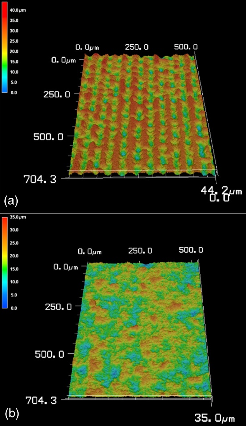
Fig. 1Mean surface roughness of ultrashort pulsed laser-ablated dentin: groups (G1 to G8) are differentiated based on the absence or presence of pulse overlap (PO; 0% or 50%, respectively) for each level of fluence used. 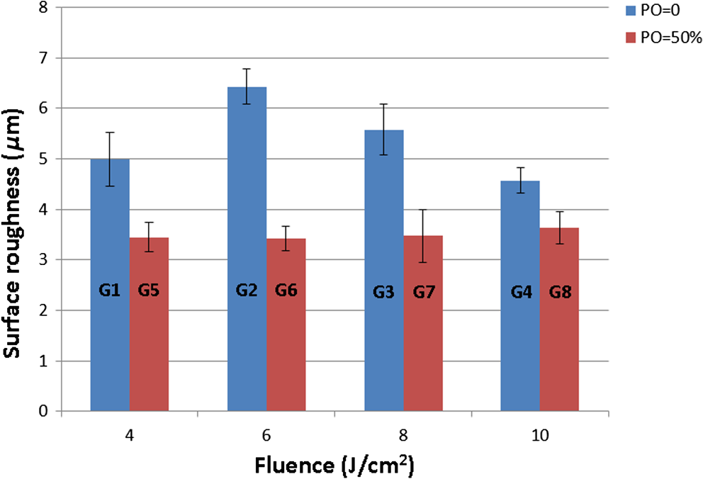 Statistical analysis of Ra was carried out among groups G1 to G4 (no PO) and G5 to G8 (50% PO) to evaluate the effect of laser fluence at the same PO. Least significant difference post-hoc comparison tests revealed that there was a significant difference between each group from G1 to G4 () and that there was no significant difference from G5 to G8 (). The influence of PO on Ra was also evaluated, albeit among group pairs (G1/G5, G2/G6, G3/G7, and G4/G8) ablated with the same laser fluence but different PO. Independent-samples tests revealed that the PO significantly influenced Ra in each pairing, regardless of fluence level (), which meant that there were significant differences between members of each group pair. Further independent-samples tests revealed that the Ra of all test groups, except for G1 and G4, was significantly different than that of control G9 (). 3.2.WettabilityAll laser-treated dentin samples showed a mean contact angle of approximately 0 deg. The mean contact angle of the control group, however, was , which was significantly different than all test groups (). 3.3.Scanning Electron Microscopy EvaluationAs shown in the micrographs of Fig. 2, following ablation, laser fluence, and PO had noticeable effects on the integrity of the dentin structure. Specifically, in G1 to G3, some of the dentin structure remained intact; clear ridges and valleys were observed, and the dentin surfaces were rougher. At the highest laser fluence (G4) or at the higher PO (G5 and G6), the dentin surfaces were entirely ablated without any remaining intact dentin structure. No smear layer was observed in any of the micrographs, and most of the dentinal tubules remained open. However, with highest laser fluence and higher PO (G8), areas of melting and resolidification could be observed. Microcracks could be observed in all groups. Fig. 2Representative micrographs of ultrashort pulsed laser-ablated dentin surfaces: (a) G1, (b) G2, (c) G3, (d) G4, (e) G5, and (f) G6. 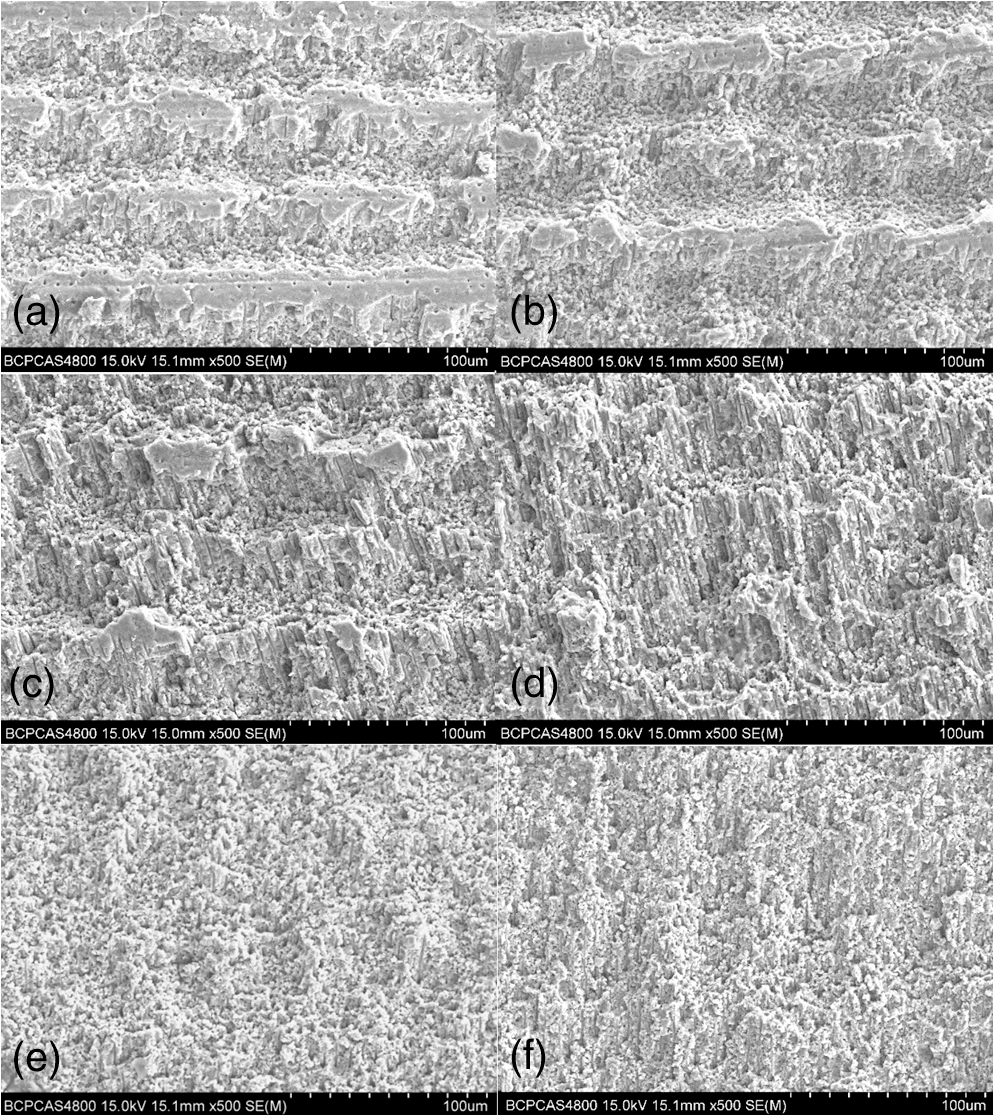 Topographic images of laser-ablated dentin were also obtained using a 3-D profile measurement laser microscope (Fig. 3). G1 topography [Fig. 3(a)] was consistent with the SEM micrograph; clear ridges and valleys were observed. G8 topography [Fig. 3(b)], which represents use of the highest laser fluence and higher PO, was also consistent with the SEM micrograph; the surface was flatter, without ridges and valleys. 4.DiscussionIn this study, different parameters of an ultrashort pulsed laser were analyzed quantitatively for their effects on the surface roughness and wettability (critical factors of bond strength) of dentin following ablation and compared to the effects of a common rotary grinding instrument. With pulse duration of 20 ps and the laser fluence, the peak power (PP) used in this study was about to . With the high PP, the effect of wavelength on dentin would be less of an issue. By reviewing the literatures, the laser fluence is considered as an important factor that influences the outcome after ablation of dental tissues,25–27 and the laser fluence also has a relationship with . The effect of PO could guide the determination of scanning speed and scanning line spacing when choosing laser parameters. It was also noted that when the highest laser fluence and higher PO were used, melting and resolidification was observed. One reason for this effect could be heat accumulation; Rego et al. reported that thermal damage could be present when higher laser energies are used or when the tooth is significantly dehydrated.28 In almost all groups, microcracks were observed. It seems that the microcracks could be induced during sample preparation for SEM evaluation. Surface roughness is an important factor that influences the bond strength between tooth structure and restorative materials. It can also have an effect on the quality of a preparation as it directly affects the accuracy of cast and prosthesis retention.29–32 The Ra value of surface roughness is the most important parameter used to describe the surface roughness of a substrate, so the current study focused on the analysis of Ra following ablation. In this study, when a PO of 0 was used, laser fluence significantly influenced dentin surface roughness, and it seems that from G2 onward, the dentin surface roughness decreased with increasing laser fluence. However, when a PO of 50% was used, laser fluence did not significantly influence dentin surface roughness. These effects can be easily reasoned. As fluence is increased, the surface is ablated with more power, thus removing more of the surface and causing it to become flatter, or less rough. When a 50% PO is added, however, each focal point is effectively ablated twice, which in this case appeared to significantly reduce the influence of laser fluence (at least at the levels tested here) on surface roughness. Notably, surface roughness of the control group (G9) appeared to be generally lower than that of some groups without PO (G1 to G3) and higher than that of groups with 50% PO (G5 to G8), albeit without statistical significance. Wettability is an important factor affecting the bond between the dental substrate and restorative materials.33,34 For having been used as a means of characterizing the wetting of a substrate by the fluid phase, contact angle measurement is considered to be a useful indicator of interfacial tension.4,35 Although it was hypothesized that surface roughness may improve wettability by reducing the contact angle,36 the mean contact angle of all laser-ablated groups in this study was approximately 0 deg, regardless of surface roughness, while that of the control group was significantly larger. Vorobyev and Guo37 reported on using a femtosecond pulse laser with a unique surface texturing approach to make human dentin superwetting. In their studies, superwetting is strongly related to the presence of open capillary systems, such as surface grooves, which depend on surface geometry.38–40 By varying the geometry of the microgrooves, wetting could be controllable. However, the femtosecond laser used in their study operated at a 1-kHz repetition rate, which was not enough to allow for efficient ablation. Some researchers have voiced concern over the low ablation efficiency of ultrashort pulsed lasers in dentistry.26,41 In recent years, however, some studies reported that, with the use of a laser scanning system, it was possible to ablate a large area with an ultrashort pulsed laser.42–44 In the current study, with the use of such a scanning system and operating at a 100-kHz repetition rate, the ablation efficiency of an ultrashort pulsed laser was indeed proven. When a lower PO was used, microgrooves were observed, possibly contributing to the superwettability of the dentin surface. When a higher PO was used, dentin surfaces were flatter and had a lower surface roughness than control group; however, superwettability still existed, which meant that microgrooves were not essential to improve the wettability of the dentin surface. The smear layer produced by mechanical rotary instruments could have a more prominent role than surface roughness in determining wettability.29,45 Indeed, it tends to act as a physical barrier to the diffusion of oral fluids and restorative materials,46 occluding dentinal tubules and reducing dentin permeability.47,48 In this study, no smear layer was observed in any of the laser-ablated dentin samples. Moreover, the dentinal tubules remained mostly open, which may have contributed to the superwettability of dentin surfaces, even of surfaces ablated with a 50% PO where the surface roughness was lower than that of the control. Achieving superwettability through ultrashort pulsed laser ablation should enhance bond strength. This effect will be explored in future studies. 5.ConclusionSurface roughness of ultrashort pulsed laser-ablated dentin decreased with higher laser fluence and PO. However, a 50% PO appeared to trump the effect of the fluence levels used in this study. Furthermore, wettability was significantly increased in all laser-ablated dentin samples, which each showed a contact angle of approximately 0 deg, compared to the manually ground control samples. AcknowledgmentsThe authors are grateful to the National Science and Technology Pillar Program during the 12th Five-Year Plan (Grant No. 2012BAI07B04) for financial support. The authors would like to thank the Institute of Physics of the Chinese Academy of Sciences for providing the laser equipment and the Oral and Maxillofacial Surgery Unit of the Peking University Hospital of Stomatology for providing the human extracted teeth used in these experiments. The authors declare no conflicts of interest. ReferencesJ. D. Eick et al.,
“Surface topography: its influence on wetting and adhesion in a dental adhesive system,”
J. Dent. Res., 51
(3), 780
–788
(1972). http://dx.doi.org/10.1177/00220345720510031401 JDREAF 0022-0345 Google Scholar
J. A. Aguilar-Mendoza et al.,
“Effect of acid etching on dentin wettability and roughness: self-etching primers versus phosphoric acid,”
J. Biomed. Mater. Res. Part B, 84
(1), 277
–285
(2008). http://dx.doi.org/10.1002/(ISSN)1552-4981 1552-4973 Google Scholar
J. D. Eick et al.,
“Current concepts on adhesion to dentin,”
Crit. Rev. Oral Biol. Med., 8
(3), 306
–335
(1997). http://dx.doi.org/10.1177/10454411970080030501 CROMEF 1045-4411 Google Scholar
W. M. Al-Omari, C. A. Mitchell and J. L. Cunningham,
“Surface roughness and wettability of enamel and dentine surfaces prepared with different dental burs,”
J. Oral Rehabil., 28
(7), 645
–650
(2001). http://dx.doi.org/10.1046/j.1365-2842.2001.00722.x JORHBY Google Scholar
H. V. Oene, Y. F. Chang and S. Newman,
“The rheology of wetting by polymer melts,”
J. Adhes., 1
(1), 54
–68
(1969). http://dx.doi.org/10.1080/00218466908077375 JATEE8 0169-4243 Google Scholar
U. Keller and R. Hibst,
“Experimental studies of the application of the Er:YAG laser on dental hard substances: II. Light microscopic and SEM investigations,”
Lasers Surg. Med., 9
(4), 345
–351
(1989). http://dx.doi.org/10.1002/(ISSN)1096-9101 LSMEDI 0196-8092 Google Scholar
R. Hibst and U. Keller,
“Experimental studies of the application of the Er:YAG laser on dental hard substances: I. Measurement of the ablation rate,”
Lasers Surg. Med., 9
(4), 338
–344
(1989). http://dx.doi.org/10.1002/(ISSN)1096-9101 LSMEDI 0196-8092 Google Scholar
A. Le Goff et al.,
“An evaluation of the CO2 laser for endodontic disinfection,”
J. Endod., 25
(2), 105
–108
(1999). http://dx.doi.org/10.1016/S0099-2399(99)80006-5 0099-2399 Google Scholar
G. B. Altshuler, A. V. Belikov and Y. A. Sinelnik,
“A laser-abrasive method for the cutting of enamel and dentin,”
Lasers Surg. Med., 28
(5), 435
–444
(2001). http://dx.doi.org/10.1002/(ISSN)1096-9101 LSMEDI 0196-8092 Google Scholar
J. Liu et al.,
“Acceptance and efficiency of Er:YAG laser for cavity preparation in children,”
Photomed. Laser Ther., 24
(4), 489
–493
(2006). http://dx.doi.org/10.1089/pho.2006.24.489 PLDHA8 1549-5418 Google Scholar
P. Ekworapoj, S. K. Sidhu and J. F. McCabe,
“Effect of different power parameters of Er, Cr:YSGG laser on human dentine,”
Lasers Med. Sci., 22
(3), 175
–182
(2007). http://dx.doi.org/10.1007/s10103-006-0426-6 LMSCEZ 1435-604X Google Scholar
S. S. Kim et al.,
“Effects of laser-irradiated dentin on shear bond strength of composite resin,”
J. Korean Acad. Prosthodontics, 46
(5), 520
–527
(2008). http://dx.doi.org/10.4047/jkap.2008.46.5.520 Google Scholar
M. Youssef et al.,
“Dentinal surface-cutting efficiency using a high-speed diamond bur, ultrasound and laser,”
Laser Phys., 18
(4), 472
–477
(2008). http://dx.doi.org/10.1134/s11490-008-4022-2 LAPHEJ 1054-660X Google Scholar
A. Almehdi et al.,
“Histological and SEM analysis of root cementum following irradiation with Er:YAG and CO2 lasers,”
Lasers Med. Sci., 28
(1), 203
–213
(2013). http://dx.doi.org/10.1007/s10103-012-1110-7 LMSCEZ 1435-604X Google Scholar
S. Shahabi, N. Chiniforush and N. Juybanpoor,
“Morphological changes of human dentin after erbium-doped yttrium aluminum garnet (Er:YAG) and carbon dioxide (CO2) laser irradiation and acid-etch technique: an scanning electron microscopic (SEM) evaluation,”
J. Lasers Med. Sci., 4
(1), 48
–52
(2013). LMSCEZ 1435-604X Google Scholar
T. Dostalova and H. Jelinkova,
“Lasers in dentistry: overview and perspectives,”
Photomed. Laser Surg., 31
(4), 147
–149
(2013). http://dx.doi.org/10.1089/pho.2013.3493 PLDHA8 1549-5418 Google Scholar
C. Momma et al.,
“Precise laser ablation with ultrashort pulses,”
Appl. Surf. Sci., 109 15
–19
(1997). http://dx.doi.org/10.1016/S0169-4332(96)00613-7 ASUSEE 0169-4332 Google Scholar
E. G. Gamaly et al.,
“Ablation of solids by femtosecond lasers: ablation mechanism and ablation thresholds for metals and dielectrics,”
Phys. Plasmas, 9
(3), 949
–958
(2002). http://dx.doi.org/10.1063/1.1447555 PHPAEN 1070-664X Google Scholar
M. H. Niemz et al.,
“Tooth ablation using a CPA-free thin disk femtosecond laser system,”
Appl. Phys. B, 79
(3), 269
–271
(2004). http://dx.doi.org/10.1007/s00340-004-1579-2 APBOEM 0946-2171 Google Scholar
A. M. Rubenchik et al.,
“Numerical simulation of ultra-short laser pulse energy deposition and bulk transport for material processing,”
Appl. Surf. Sci., 127 193
–198
(1998). http://dx.doi.org/10.1016/S0169-4332(97)00631-4 ASUSEE 0169-4332 Google Scholar
F. Schelle et al.,
“Transmission of 1064 nm laser radiation during ablation with an ultra-short pulse laser (USPL) system,”
Proc. SPIE, 8208 82080J
(2012). http://dx.doi.org/10.1117/12.910180 PSISDG 0277-786X Google Scholar
M. C. Luengo et al.,
“Evaluation of micromorphological changes in tooth enamel after mechanical and ultrafast laser preparation of surface cavities,”
Lasers Med. Sci., 28
(1), 267
–273
(2013). http://dx.doi.org/10.1007/s10103-012-1144-x LMSCEZ 1435-604X Google Scholar
L. Ji et al.,
“Ti:sapphire femtosecond laser ablation of dental enamel, dentine, and cementum,”
Lasers Med. Sci., 27
(1), 197
–204
(2012). http://dx.doi.org/10.1007/s10103-011-0932-z LMSCEZ 1435-604X Google Scholar
M. M. Portillo et al.,
“Morphological alterations in dentine after mechanical treatment and ultrashort pulse laser irradiation,”
Lasers Med. Sci., 27
(1), 53
–58
(2012). http://dx.doi.org/10.1007/s10103-010-0845-2 LMSCEZ 1435-604X Google Scholar
P. Kohns, P. Zhou and R. S. O Rmann,
“Effective laser ablation of enamel and dentine without thermal side effects,”
J. Laser Appl., 9 171
(1997). http://dx.doi.org/10.2351/1.4745457 JLAPEN 1042-346X Google Scholar
J. K. U. Ger, W. Kautek and H. Newesely,
“Femtosecond-pulse laser ablation of dental hydroxyapatite and single-crystalline fluoroapatite,”
Appl. Phys. A, 69
(7), 403
–407
(1999). http://dx.doi.org/10.1007/s003390051426 APAMFC 0947-8396 Google Scholar
B. Kim et al.,
“Effects of high repetition rate and beam size on hard tissue damage due to subpicosecond laser pulses,”
Appl. Phys. Lett., 76
(26), 4001
–4003
(2000). http://dx.doi.org/10.1063/1.126847 APPLAB 0003-6951 Google Scholar
F. F. A. Rego et al.,
“Influence of the hydration state on the ultrashort laser ablation of dental hard tissues,”
Lasers Med. Sci., 28
(1), 215
–222
(2013). http://dx.doi.org/10.1007/s10103-012-1118-z LMSCEZ 1435-604X Google Scholar
M. F. Ayad et al.,
“Influence of dental rotary instruments on the roughness and wettability of human dentin surfaces,”
J. Prosthet. Dent., 102
(2), 81
(2009). http://dx.doi.org/10.1016/S0022-3913(09)60114-1 JPDEAT 0022-3913 Google Scholar
M. F. Ayad, S. F. Rosenstiel and M. M. Hassan,
“Surface roughness of dentin after tooth preparation with different rotary instrumentation,”
J. Prosthet. Dent., 75
(2), 122
–128
(1996). http://dx.doi.org/10.1016/S0022-3913(96)90087-6 JPDEAT 0022-3913 Google Scholar
M. F. Ayad, S. F. Rosenstiel and M. Salama,
“Influence of tooth surface roughness and type of cement on retention of complete cast crowns,”
J. Prosthet. Dent., 77
(2), 116
–121
(1997). http://dx.doi.org/10.1016/S0022-3913(97)70223-3 JPDEAT 0022-3913 Google Scholar
D. Chan et al.,
“Effect of preparation convergence on retention and seating discrepancy of complete veneer crowns,”
J. Oral Rehabil., 32
(1), 58
–64
(2005). JORHBY Google Scholar
C. T. Preoteasa et al.,
“Wettability of some dental materials,”
Optoelectron. Adv. Mater. Rapid Commun., 5
(8), 874
–878
(2011). 1454-4164 Google Scholar
D. Munirathinam, D. Mohanaj and M. Beganam,
“Efficacy of various cleansing techniques on dentin wettability and its influence on shear bond strength of a resin luting agent,”
J. Adv. Prosthodontics, 4
(3), 139
–145
(2012). http://dx.doi.org/10.4047/jap.2012.4.3.139 Google Scholar
B. W. Darvell, M. D. Murray and N. H. Ladizesky,
“Contact angles: a note,”
J. Dent., 15
(2), 82
–84
(1987). http://dx.doi.org/10.1016/0300-5712(87)90005-4 JDENAB 0300-5712 Google Scholar
M. Toledano et al.,
“Effect of acid etching and collagen removal on dentin wettability and roughness,”
J. Biomed. Mater. Res., 47
(2), 198
–203
(1999). http://dx.doi.org/10.1002/(ISSN)1097-4636 JBMRBG 0021-9304 Google Scholar
A. Y. Vorobyev and C. Guo,
“Making human enamel and dentin surfaces superwetting for enhanced adhesion,”
Appl. Phys. Lett., 99
(19), 193703
(2011). http://dx.doi.org/10.1063/1.3660579 APPLAB 0003-6951 Google Scholar
A. Y. Vorobyev and C. Guo,
“Metal pumps liquid uphill,”
Appl. Phys. Lett., 94
(22), 224102
(2009). http://dx.doi.org/10.1063/1.3117237 APPLAB 0003-6951 Google Scholar
A. Y. Vorobyev and C. Guo,
“Water sprints uphill on glass,”
J. Appl. Phys., 108
(12), 123512
(2010). http://dx.doi.org/10.1063/1.3511431 JAPIAU 0021-8979 Google Scholar
A. Y. Vorobyev and C. Guo,
“Laser turns silicon superwicking,”
Opt. Express, 18
(7), 6455
–6460
(2010). http://dx.doi.org/10.1364/OE.18.006455 OPEXFF 1094-4087 Google Scholar
M. H. Niemz and J. Neev,
“Ultrashort laser pulses in dentistry: advantages and limitations,”
Proc. SPIE, 3255 84
–91
(1998). http://dx.doi.org/10.1117/12.308211 Google Scholar
J. Serbin et al.,
“Femtosecond lasers as novel tool in dental surgery,”
Appl. Surf. Sci., 197 737
–740
(2002). http://dx.doi.org/10.1016/S0169-4332(02)00402-6 ASUSEE 0169-4332 Google Scholar
M. Straßl et al.,
“Novel applications of short and ultra-short pulses,”
Appl. Surf. Sci., 247
(1), 561
–570
(2005). http://dx.doi.org/10.1016/j.apsusc.2005.01.174 ASUSEE 0169-4332 Google Scholar
M. Straßl, A. Yousif and E. Wintner,
“Scanning of ultra-short laser pulses in dental applications. A comparison of scanning algorithms and pulse durations,”
J. Oral Laser Appl., 7
(2), 123
–128
(2007). 1473-7809 Google Scholar
J. D. Eick et al.,
“Scanning electron microscopy of cut tooth surfaces and identification of debris by use of the electron microprobe,”
J. Dent. Res., 49
(6), 1359
–1368
(1970). http://dx.doi.org/10.1177/00220345700490063601 JDREAF 0022-0345 Google Scholar
D. H. Pashley et al.,
“Scanning electron microscopy of the substructure of smear layers in human dentine,”
Arch. Oral Biol., 33
(4), 265
–270
(1988). http://dx.doi.org/10.1016/0003-9969(88)90188-4 AOBIAR 0003-9969 Google Scholar
J. A. Barros et al.,
“Effect of bur type and conditioning on the surface and interface of dentine,”
J. Oral Rehabil., 32
(11), 849
–856
(2005). http://dx.doi.org/10.1111/j.1365-2842.2005.01507.x JORHBY Google Scholar
J. Tagami et al.,
“Effects of high-speed cutting on dentin permeability and bonding,”
Dent. Mater., 7
(4), 234
–239
(1991). http://dx.doi.org/10.1016/S0109-5641(05)80021-1 DEMAEP 0109-5641 Google Scholar
BiographyJing Liu received her BS degree at the China Medical University, Shenyang, China, in 2011. She is now a PhD student at Peking University School and Hospital of Stomatology, Beijing, China. Her research focuses on laser effects on dental tissues. Peijun Lü obtained his PhD degree in medicine in 1994 and is affiliated with the Department of Prosthodontics, Peking University School of Stomatology, where he has worked as a professor and chief physician for more than 15 years. He has also been vice director of the National Engineering Laboratory for Digital and Material Technology of Stomatology since 2011. His research interesting is in digital and material technology pertinent to stomatology. Yuchun Sun received his DDS degree in prosthodontics from Peking University in 2009. He has worked in the research fields of dental clinic treatments, three-dimensional (3-D) digital dental technology, and dental material for over 10 years at the Peking University School and Hospital of Stomatology. His current research interests concern digital technologies in laser dentistry. Yong Wang received his MS degree in engineering mechanics from Peking University in Beijing, China, in 1988. He has been principle engineer (professor), deputy director, of the Center of Digital Dentistry at Peking University School and Hospital of Stomatology for more than 15 years. His interesting research fields include analysis of 3-D data in dentistry, cross-subjects of dental clinical trials, and digital techniques. |
||||||||||||||||||||||||||||||||||||||||||||||||||||||||||||||||||||||