
|
|
|
In past decades, diffuse optical imaging (DOI) has been an emerging imaging modality, probing biological tissue by using near-infrared spectroscopy (NIRS), which shows the abilities of noninvasive detection at high temporal resolution as well as molecular function imaging with high specificity and sensitivity in human tissue.1–3 However, to focus on imaging, most biological tissues are highly scattering and, therefore, the incident photons are quickly diffused before being absorbed or detected. Therefore, imaging by using NIR light becomes necessary because of its greater transparency in human tissue compared with the visible light.1 Experimentally, measuring amplitude and phase of diffuse photon density wave (DPDW) can recover the image object in a multiple scattering medium via the distribution of position-dependent absorption and reduced scattering coefficients. However, this is based on the simplified diffusion equation, where a position-independent diffusion coefficient is assumed.4–6 The constant diffusion coefficient in the diffusion equation enables smoothing out the boundary or surface effect of a small object via an inverse algorithm in the recovery image. In addition, DPDW is generated in the scattering media by using a high-frequency intensity-modulation light source that produces high-level radio frequency (RF) intensity noise in the detected intensity-modulation signal. Then, lower spatial resolution in the recovery image results apparently.4–7 In order to improve the spatial resolution of the image, the boundary or surface effect of a small scattering object in a multiple scattering medium becomes critical according to the perturbation theory derived by Ostermeyer and Jacques.4,5 Consequently, the properties of the signal induced by the boundaries of scattering objects in multiple scattering media are examined in this study. Boas et al.6 derived the perturbation theory of scattered DPDW by a spherical inhomogeneity in turbid media, where a time-dependent solution of the Helmholtz equation was achieved by using a sinusoidally intensity-modulated point source in a multiple scattering medium under the condition of uniform distribution of diffusion coefficient.6–8 The amplitude and phase of the scattered DPDW induced by a spherical inhomogeneity were analytically calculated and experimentally verified.6–9 In contrast, Ostermeyer and Jacques4 derived an analytical model of solving the diffusion equation of inhomogeneous diffusion coefficient using perturbation theory. When only sharply bounded inhomogeneity with constant optical properties inside is considered, the fluence perturbation induced by an object in scattering media can be divided into two parts: (1) the surface effect and (2) the volume effect. For the absorber, the contribution from the surface effect is less important than for the scattering effect and vice versa for the scatter in scattering media.4 Meanwhile, in the case of a small object by the surface-to-volume ratio, , the surface effect becomes dominant in perturbation theory, and then approaches a dipole field. Clinically, the tumor as an image object in tissue generally presents a sharper boundary than a normal cell at early stage.1,10,11 Meanwhile, the refractive index mismatch introduces scattering effect at the boundary, which enhances the surface effect. The surface effect relating to the boundary of the small object dominates the spatial resolution of the recovered image, whereas the volume effect smoothes out of the structure image by volume integration.4 As a result, the capability of detecting the surface effect from a small object via the amplitude and phase change detection of diffuse photon-pair density wave (DPPDW) has the potential to increase the resolution of the inverse problem not only by direct detection of the surface effect by a small object but also the higher detection sensitivity of the heterodyne signal versus the polarization gating and coherence gating of scattered linear-polarized photon pairs (LPPP).12–14 However, there has been no experimental demonstration of the surface effect of a small object in a multiple scattering medium, being made possible by means of the conventional DPDW method. One of the reasons is due to the large amount of RF noise generated by a high-frequency intensity modulation of the light source being required in the DPDW setup. As a result, the sensitivities as well as the spatial resolution of the amplitude and phase measurements of the scattered DPDW close to the boundary of the object are limited in the experiment. In our previous studies, the properties of DPPDW in turbid media have been studied.12–17 This is based on the features of two-frequency linear-polarized highly correlated photon-pairs laser beam propagating in turbid media via the coherence and polarization gatings, simultaneously, and the heterodyne synchronized detection.12–17 Theoretically, the surface effect, which is induced by the boundary of a small scattering object following a dipole-like perturbation,4 is anticipated both in amplitude and phase of scattered DPPDW. They are localized close to the boundaries of the image object because DPPDW satisfies the diffusion equation. 12–14 In this experiment, we measured the surface effect of a 2-mm (in diameter) spherical scatter in 1% intralipid solution. The experimental result agrees well with the theoretical calculations following the perturbation theory derived by Ostermeyer and Jacques. According to our developed theory, DPPDW provides a coherent detection of photon density wave, which is produced by the collection of the scattered LPPP in a highly scattering medium. LPPP comprises a pair of linearly parallel-polarized light waves with high-spatial and -temporal coherence. The pair of photons also presents a slight difference in temporal frequency derived from a two-frequency laser. Generally, LPPP can be detected via the heterodyne interference signal between the pair of linear-polarized waves and is expressed as where and are the amplitudes of the paired linear-polarized photons with the corresponding frequencies and , respectively. is the beat frequency of the two waves, and is the path-dependence phase difference between the pair of linear-polarized photons, where is the optical path length and is the light speed in the medium. The term represents the heterodyne efficiency. As the detection signal of LPPP depends on the amplitude modulation, this ensures the features of high signal-to-noise ratio (SNR), narrower detection bandwidth, and wider dynamic range compared to the intensity modulation adapted by DPDW in the conventional frequency-domain diffusion method. According to the theory of DPPDW, the propagation of LPPP in a highly scattering medium satisfies the diffusion equation. Thus, in a homogeneous and infinite highly scattering medium, DPPDW is written as12–14 where is the distance between the emitter and the receiver. and are the real part and the imaginary part of the wave number of DPPDW, respectively, and are expressed by and where and are the absorption coefficient and the reduced scattering coefficient of the scattering medium, respectively. The phase velocity of the DPPDW is expressed by Equations (2)–(5) represent the optical properties of DPPDW with paired highly correlated and polarized photons. Compared with the DPDW, the imaginary part of the complex wavenumber of the DPPDW () is linearly proportional to the beat frequency and the real part of the complex wavenumber () is independent of . However, the real part and the imaginary part of the wavenumber of the DPDW, and are both proportional to the square root of the intensity-modulated frequency () in the high-frequency regime.8 The phase velocity of the DPPDW is similar to that of a DPDW at a lower modulation frequency, whereas the phase velocity of the DPPDW is different from that of a DPDW at a higher modulation frequency (), where the phase velocity of DPDW depends on .In a highly scattering medium, however, the image quality depends on the degree of coherence and degree of polarization of scattered LPPP. This means that the detection of scattered LPPPs in the scattering medium is filtered by the coherence gating, the polarization gating, and, finally, the electronic filter gating via a lock-in amplifier.12–17 Those features are introduced by the coherence and polarization properties of DPPDW via heterodyning. In order to verify the ability to detect the surface effect of a small object, the amplitude and phase responses of DPPDW close to the boundary of the scatter were measured. Meanwhile, to validate the experimental results, the amplitude and phase responses of the surface effect by DPPDW are calculated by adapting the complex Green’s function of DPPDW into the perturbation theory.4 Figure 1 shows the experimental setup wherein a two-frequency laser (ZMI 7702 laser head, Zygo) was used, which provides the LPPP laser beam by use of an analyzer at azimuth angle of 45 deg to the -axis located in front of the laser. LPPP requires a pair of parallel linearly polarized light waves with a slight difference (20 MHz) in temporal frequency. The output power and center wavelength of this laser beam are 0.6 mW and 632.8 nm, respectively. A water tank filled with 1% intralipid solution was taken as an infinite and homogeneous multiple scattering medium. This gave a reduced scattering coefficient and an absorption coefficient . The laser beam is delivered into the scattering medium through a source fiber, whereas the fluence of scattered LPPPs is collected by a movable detector fiber and detected by a photomultiplier tube. Finally, the amplitude and phase signals of DPPDW are measured simultaneously by use of a lock-in amplifier. In this experiment, a scattering sphere with a 2-mm diameter was made with optical properties and from a polyester resin mixture containing 3.5-mg powder (scatters) for every 1 ml of polyester resin.18 There is no extra absorption dye in the resin mixture. The scattering order of the scatter to the background medium by was 0.3. The geometry of measurement is shown in Fig. 2, where the source fiber was fixed at , and the sphere was fixed at in the tank. For the detection of surface effect, the total complex fluence (i.e., amplitude and phase) of DPPDW () was measured as a function of the position of the detector fiber, which was scanned from to at 1-mm intervals. The moving step was adjusted to 0.5 mm when the detector fiber was moving close to the scattered sphere to ensure that the surface effect was measured properly. In addition, the homogeneous complex fluence () was also measured under the same arrangement without the scattering sphere. Thus, the perturbation complex fluence or the surface effect can be obtained by subtracting from . Note that the size of the sphere is small enough that it satisfies the condition of surface-to-volume ratio . Then, is dominated by the surface effect and the volume effect is ignored. Fig. 1The experimental setup of amplitude and phase detection of DPPDW for the measurement of the surface effect of a scattering sphere in a highly scattering medium. 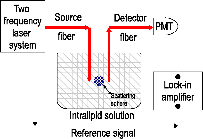 Fig. 2Schematic geometry of surface effect measurement for a scattering sphere in a multiple scattering medium. The source fiber was fixed at and the sphere was fixed at in the tank. The detector fiber was scanned along -axis to collect the total complex fluence of DPPDW.  Figure 3 shows the experimental results (dots) of (a) the amplitude and (b) the phase response of the surface effect measured by DPPDW. Both results are compared to the theoretical calculation (solid curves) according to the perturbation theory.4 In this theoretical calculation, and of this scattering sphere (2 mm in diameter) are 28 and , respectively; and of the turbid medium are 14 and , respectively. In Fig. 3(a), the amplitude response was normalized by and the surface effect, which produces a rapid change close to the boundary of the scattering object, is seen clearly. This result shows a dipole-like perturbation whereas the magnitude is positive at the front surface () and then becomes negative at the rear surface () of the scattered sphere. Note that the dipole-like perturbation is asymmetric and the magnitude of the positive peak is larger than that of the negative one. These results agree well with prediction from perturbation theory (), where the volume effect is ignored. The phase signal of DPPDW is also shown in Fig. 3(b) and the result is similar to the amplitude response (). These results not only show high sensitivity to surface effect detection but also no surface effect mismatch following the prediction of perturbation theory4 under a strong surface effect by a small scattering object in turbid media. This also implies a significant improvement on spatial resolution of the image of a small object in turbid media. Fig. 3The surface effect of a 2-mm-in-diameter scattering sphere in (a) amplitude response and (b) phase response measured by the DPPDW method. Both present the dipole-like perturbation with a rapid change close to the scatter boundary. The correlation between measured data with prediction are in (a) and in (b). In homogeneous multiple scattering media, the amplitude and phase responses of DPPDW in setup without scattering object in 1% intralipid solution is shown in (c).  From these experimental results, we also validate that the propagation of DPPDW satisfies the diffusion equation in terms of perturbation theory of position-dependent diffusion coefficient near image object in a multiple scattering medium. Meanwhile, using DPPDW, high-detection sensitivity at a relatively low beat frequency (20 MHz) is applicable as well. In this experiment, the surface effect of the 2-mm scatter in 1% intralipid solution are in amplitude perturbation and in phase perturbation close to the boundary of the scatter. This, for the first time, shows the surface effect of a small scatter (2 mm in diameter), which leads to rapid changes of polarized photon-pairs fluence both in amplitude and phase at the boundary of the scatter. The perturbation theory of diffusion equation with position-dependent diffusion coefficient in frequency domain has also been verified. It is in contrast to the conventional DPDW method wherein the surface effect is not available due to high levels of RF noise inherent with high-frequency modulation. From this result, DPPDW potentially becomes an approach to improve the spatial resolution of the recovery image in multiple scattering media in comparison with DPDW. These results not only enhance the boundary detection sensitivity of a scatter but also provide the possibility of enhancing the boundary detection sensitivity of an absorber via its phase response. This is because the phase difference of DPPDW close to the boundary of an absorber is dependent. (The phase of DPPDW is proportional to which is dependent, so the sensitivity of phase change across the boundary of an absorber in a multiple scattering medium becomes dependent.) As a result, the localization of surface boundary of an absorber in multiple scattering media can be enhanced at lower via DPPDW accordingly. In contrast, the features of coherence and polarization gatings of LPPPs in DPPDW method mean that the common background phase noise is reduced significantly due to the common-path propagation of LPPP in turbid media.12–17 This also introduces less dephasing or higher SNR in the detected beat signal that is beneficial to surface effect detection sensitivity. Additionally, the amplitude response via heterodyne interference in DPPDW also increases the dynamic range of detection in contrast to the high-frequency intensity-modulated signal in a conventional DPDW setup. Clinically, these advantages for surface effect detection can become very critical, e.g., to differentiate a benign tumor from a malignant one due to the surface scattering effect being dominant in a malignant tumor.10 In addition, a nonpolarized photon-pair laser beam can be used in the DPPDW method, in which highly spatial and temporal correlated but nonpolarized photon pairs propagate in a scattering medium. The advantage of using nonpolarized photon pairs is that DPPDW becomes insensitive to birefringent properties of the scattering medium. However, using a linear-polarized photon-pairs laser beam could introduce polarized image contrast and maybe further improve the quality of recovered images. In conclusion, the capability of measuring the surface effect of a small scattering object in a multiple scattering medium via DPPDW has been experimentally verified. As the surface effect presents a sharply dipole-like amplitude and phase change of DPPDW near the boundary of a small scattering object in turbid media, it becomes highly sensitive to the shape and orientation of the image object. Therefore, we can anticipate the ability to localize the boundary of scattering objects in turbid media via the surface effect that is potentially able to improve the spatial resolution in the recovered image. With regards to an absorber in turbid media, the phase response of DPPDW can also be used to enhance the boundary detection sensitivity of an absorber due to dependence in the phase response, particularly for the image object with lower . Furthermore, polarized or nonpolarized photon pairs can be used as a light source in the DPPDW method. AcknowledgmentsThis research was supported by the Ministry of Science and Technology (MOST) of Taiwan. ReferencesT. Durduran et al.,
“Diffuse optics for tissue monitoring and tomography,”
Rep. Prog. Phys., 73 076701
(2010). http://dx.doi.org/10.1088/0034-4885/73/7/076701 RPPHAG 0034-4885 Google Scholar
V. Ntziachristos and B. Chance,
“Breast imaging technology: probing physiology and molecular function using optical imaging—applications to breast cancer,”
Breast Cancer Res., 3
(1), 41
–46
(2001). http://dx.doi.org/10.1186/bcr269 BCTRD6 Google Scholar
D. A. Boas et al.,
“The accuracy of near infrared spectroscopy and imaging during focal changes in cerebral hemodynamics,”
NeuroImage, 13
(1), 76
–90
(2001). http://dx.doi.org/10.1006/nimg.2000.0674 NEIMEF 1053-8119 Google Scholar
M. Ostermeyer and S. Jacques,
“Perturbation theory for diffuse light transport in complex biological tissues,”
J. Opt. Soc. Am. A, 14
(1), 255
–261
(1997). http://dx.doi.org/10.1364/JOSAA.14.000255 JOAOD6 0740-3232 Google Scholar
S. Feng, F. A. Zeng and B. Chance,
“Photon migration in the presence of a single defect: a perturbation analysis,”
Appl. Opt., 34
(19), 3826
–2837
(1995). http://dx.doi.org/10.1364/AO.34.003826 APOPAI 0003-6935 Google Scholar
D. Boas et al.,
“Scattering of diffuse photon density waves by spherical inhomogeneities within turbid media: analytical solution and applications,”
Proc. Natl. Acad. Sci. U. S. A., 91
(11), 4887
–4891
(1994). http://dx.doi.org/10.1073/pnas.91.11.4887 Google Scholar
M. A. O’Leary et al.,
“Experimental images of heterogeneous turbid media by frequency-domain diffusing photon tomography,”
Opt. Lett., 20 426
–428
(1995). http://dx.doi.org/10.1364/OL.20.000426 OPLEDP 0146-9592 Google Scholar
B. J. Tromberg et al.,
“Properties of photon density waves in multiple-scattering media,”
Appl. Opt., 32
(4), 607
–616
(1993). http://dx.doi.org/10.1364/AO.32.000607 APOPAI 0003-6935 Google Scholar
J. B. Fishkin and E. Gratton,
“Propagation of photon density wave in strongly scattering media containing an absorbing semi-infinite plane bounded by a straight edge,”
J. Opt. Soc. Am. A, 10
(1), 127
–140
(1993). http://dx.doi.org/10.1364/JOSAA.10.000127 JOAOD6 0740-3232 Google Scholar
D. R. Leff et al.,
“Diffuse optical imaging of the healthy and diseased breast: a systematic review,”
Breast Cancer Res. Treat., 108
(1), 9
–22
(2008). http://dx.doi.org/10.1007/s10549-007-9582-z BCTRD6 Google Scholar
B. Beauvoit et al.,
“Correlation between the light scattering and the mitochondrial content of normal tissues and transplantable rodent tumors,”
Anal. Biochem., 226
(1), 167
–174
(1995). http://dx.doi.org/10.1006/abio.1995.1205 ANBCA2 0003-2697 Google Scholar
Y. H. Chan et al.,
“Properties of a diffused photon-pair density wave in a multiple-scattering medium,”
Appl. Opt., 44
(8), 1416
–1425
(2005). http://dx.doi.org/10.1364/AO.44.001416 APOPAI 0003-6935 Google Scholar
L. P. Yu et al.,
“Detection and characterization of an optical inhomogeneity by diffuse photon-pairs density wave in a multiple-scattering medium,”
Proc. SPIE, 6629 66290F
(2007). http://dx.doi.org/10.1117/12.728617 PSISDG 0277-786X Google Scholar
L. P. Yu et al.,
“Measurement of diffuse photon-pairs density wave in a multiple-scattering medium,”
Appl. Opt., 47
(14), 2708
–2714
(2008). http://dx.doi.org/10.1364/AO.47.002708 APOPAI 0003-6935 Google Scholar
C. Chou et al.,
“Polarized optical coherence imaging in turbid media by use of a Zeeman laser,”
Opt. Lett., 25
(20), 1517
–1519
(2000). http://dx.doi.org/10.1364/OL.25.001517 OPLEDP 0146-9592 Google Scholar
L. C. Peng et al.,
“Zeeman laser-scanning confocal microscopy in turbid media,”
Opt. Lett., 26
(6), 349
–351
(2001). http://dx.doi.org/10.1364/OL.26.000349 OPLEDP 0146-9592 Google Scholar
L. P. Yu et al.,
“Glucose detection in a highly scattering medium with diffuse photon-pair density wave,”
J. Innovative Opt. Health Sci., 10
(1), 1650032
(2016). http://dx.doi.org/10.1142/S1793545816500322 Google Scholar
M. Firbank and D. T. Delpy,
“A design for a stable and reproducible phantom for use in near infra-red imaging and spectroscopy,”
Phys. Med. Biol., 38
(6), 847
–853
(1993). http://dx.doi.org/10.1088/0031-9155/38/6/015 PHMBA7 0031-9155 Google Scholar
|


