
|
|
1.IntroductionThe fundamental role of water in the functioning of living systems is well known. Even a minor violation of the water balance (by 10%) may lead to a development of pathological conditions in these systems. The main part of water in an organism is bound by connective tissues that are able to retain water due to the presence of glycosaminoglycans in the intercellular matrix.1,2 The corneoscleral shell of the eye, being a connective tissue formation, normally contains a significant quantity of water. For example, the normal human cornea contains about 78% of water mass while the normal sclera has 65%.3 If excessive water is accumulated in the cornea (the corneal edema), the tissue’s refractive properties are changing; especially important is the fact that the cornea loses its transparence, which leads to significant loss of vision. On the other hand, a dehydrated cornea also changes its shape and refractive ability; if the loss of water persists, the cornea is subject to a dystrophic process that causes irreversible violations of visual function.4 Violated water balance of the cornea may be due to a variety of eye diseases (inflammatory processes, injury, and corneal ectasias) as well as a number of medical activities, including surgical interventions such as keratorefractive operations for myopia and other refraction abnormalities, cross-linking for keratoconus, therapeutic procedures, such as prolonged instillation of certain medications (e.g., hypotensive ones), and optical techniques (contact lens wearing).5 The sclera of the eye, which accomplishes the supportive function for the internal shells (the choroid and the retina) and other eye structures, plays an important role in the maintenance of the shape of the eye, while the violation of its biomechanical properties caused by the development of a dystrophic process in the scleral tissue is the leading cause of myopia progression and the onset of disabling complications.6–8 In addition, the changed structure and properties of the corneoscleral shell of the eye have recently been found to be a significant factor in glaucoma development.9–11 It is therefore understandable that being able to determine the extent of hydration of the cornea and the sclera may have a significant impact for diagnosing eye diseases. In view of the above, adequate control of corneal and scleral hydration is very important for early diagnosis of a variety of eye diseases (corneal dystrophies, keratoconus, progressing myopia, and open-angle glaucoma), stating indications for and contraindications against keratorefractive surgeries, the choice of effective and safe plans of local medicament treatment, including tear replacement and hypotensive therapy, as well as the choice of contact lens correction solutions. Yet, no such method is available for a clinical ophthalmologist so far. Using THz radiation (in 0.3- to 3-THz frequency range) for these purposes appears to be very promising, as it is known that, in this frequency range, water shows high absorption values and dielectric permeability, which determines a high value of the reflection coefficient. This opens up vistas for using THz scanning in the light reflected from material surfaces, including those of biological materials, to detect even the slightest changes in the concentration of water contained therein. Only a few research groups the world over work at creating systems of THz scanning of hydration in biological tissues. These groups mainly use pulsed sources of THz. To give an example, a paper of Tewari et al.12 presents a THz scanning method of porcine cornea hydration. The authors used a traditional method of obtaining pulse THz radiation, involving a photoconductive antenna and a pumping femtosecond laser. They used a detector based on Schottky barrier diode as a receiver. This scheme may be replaced by a fast and more sensitive detector based on hot electron bolometer.13 A similar technique was used previously by other authors; in particular, the paper of Taylor et al.14 presents a THz system of creating images that permit differentiating, with high precision, tissue areas with different water contents. Other authors report on a successful application of THz diagnosing of skin carcinoma15 and melanoma, as well as the analysis of the state of skin burns based on water concentration measurements.16,17 The paper of Bennett et al.18 presents the results of measuring the coefficient of reflection of porcine cornea depending on its hydration, within the 0.21-THz frequency range. The authors of this paper revealed an approximate linear dependence of the reflection coefficient on water concentration of the tissue, with a monotonically reducing slope coefficient and radiation frequency growth. In the paper of Taylor et al.,19 the results of in vivo measuring of the rabbit cornea reflectance using a pulsed THz imaging system (0.47 to 0.58 THz) and millimeter (MM)-wave reflectometer (100 GHz) are presented. The positive correlations were established between the measurements of central cornea thickness and MM-wave reflectivity. However, the technique used in the abovementioned papers is still far from implementation in practical devices. This is explained by the fact that to realize the suggested scheme of generation of THz radiation, a powerful, large, and extremely expensive femtosecond laser is needed. In addition, spectra of reflectance and transmittance of the tissue in MM-wave range () have not been presented in the papers described above. The present paper reports the results of a study of transmittance and reflectance of the cornea and the sclera in a THz range (0.13 to 0.32 THz). The radiation sources we used were inexpensive and easy-to-use generators based on a backward-wave oscillator (BWO) and impact ionization avalanche transit-time diode, while the detectors were diodes with Schottky barriers and a Golay cell. The equipment used will allow us to create a simple and a relatively inexpensive device for noninvasive control of water content in eye shells, suitable for broad practical application. 2.ExperimentThe experiments were performed on three corneas, three rabbit scleras, two rabbit eyes, and three human scleras. This study was approved by the local ethics committee of the Moscow Helmholtz Research Institute of Eye Diseases. All the experiments performed in this study have been carried out according to the World Medical Association statement on animal use in biomedical research. The experiments were undertaken according to the ethical policy of the Moscow Helmholtz Research Institute of Eye Diseases. Photos of the samples are presented in Fig. 1. We used the following experimental setup for measurements of the frequency dependence of the corneal and scleral transmittance in the sub-THz range. We employed a tunable generator in the range of 129.2 to 145.5 GHz with the step of about 10 MHz determined by the effective bandwidth of generated frequency as a radiation source. The output power was amplitude modulated by an external generator with the frequency of 10 Hz. The experimental tissues were placed into a Petri dish with a bottom about 0.8-mm thick. The Petri dish was placed at the focus of the optical system, which consisted of two off-axis parabolic mirrors with the focal distances of 50.8 and 152.4 mm, respectively. The effective beam diameter at the focus was . We used a Golay cell as a detector of the transmitted radiation. The electrical signal from the detector was measured by a lock-in amplifier. The reference signal was fed to the lock-in amplifier from an external low-frequency generator. The block diagram of the experimental setup is shown in Fig. 2. It should be noted that the output power of the generator strongly depended on frequency. For this reason, we measured the transmittance of the optical path with the Petri dish before the experiment. The transmittance spectra of the investigated tissues were obtained by dividing the registered signal at each frequency by the corresponding value of the signal obtained during calibration. Fig. 2Block diagram of the experimental setup for measurements of corneal and scleral transmittance in sub-THz frequency range. 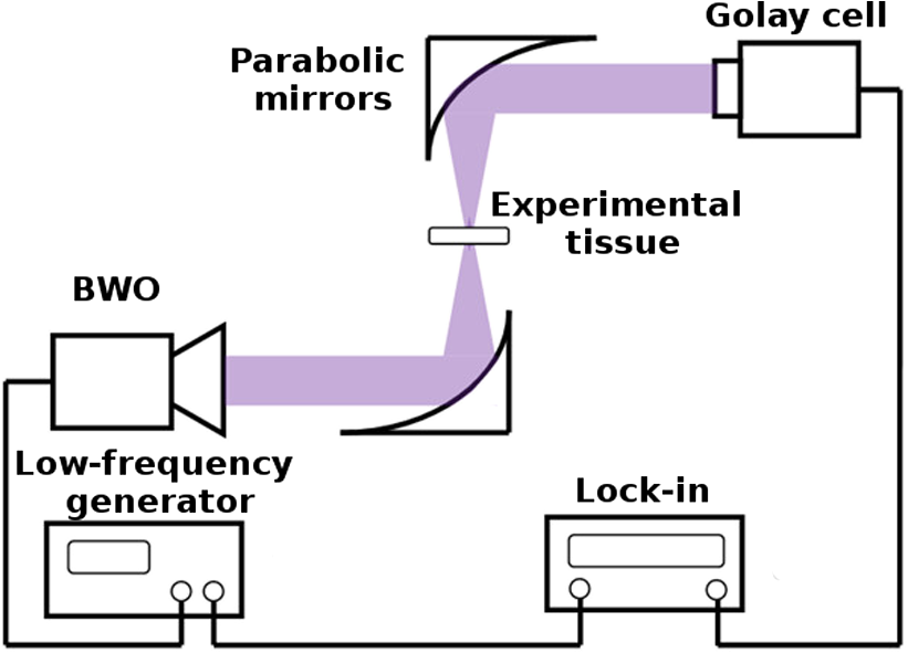 The experimental setup for measuring the reflectance coefficient depending on the mass of water content in the tissue is shown in Fig. 3. As a source of radiation, we used an avalanche transit-time diode (ATTD) with the generated frequency of 95 GHz. The generated radiation was transmitted to the sample under investigation, located in the Petri dish, using a special horn. The output power was amplitude modulated at the frequency of 16.6 kHz by an internal generator of the ATTD power supply unit. We used a detector based on a Schottky barrier diode to measure the reflected radiation. The electrical signal from the detector was measured by a lock-in amplifier. The reference signal was fed to the lock-in amplifier from the electrical low-frequency generator of ATTD power supply unit. We used a second horn to match the radiation reflected from the tissue under investigation with the detector. Fig. 3Block diagram of the experimental setup for measurements of the dependence of the corneal and scleral reflectance in the sub-THz range on the water content. 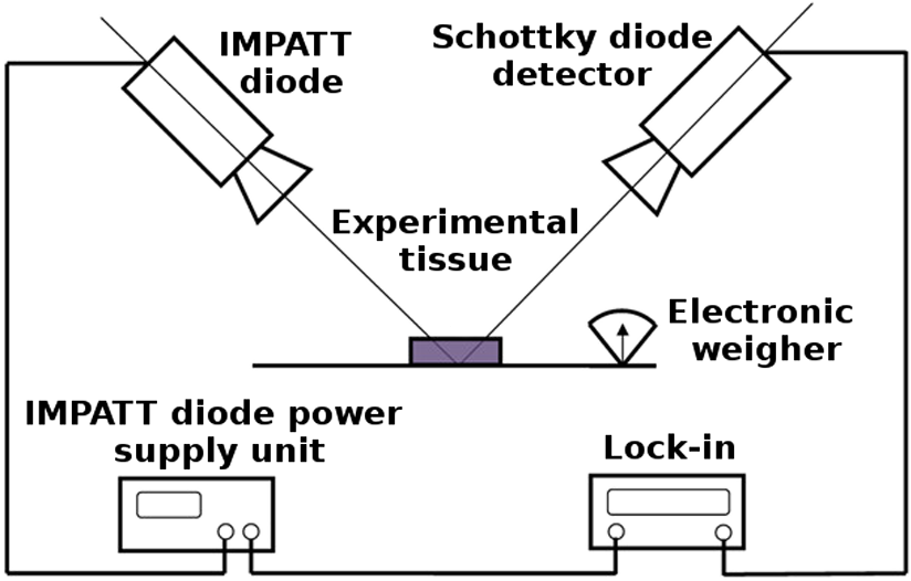 The measurement of the tissue mass was performed using electronic scales with an accuracy of g on which the Petri dish was installed. We measured the dependence of the reflected signal on the water content in the rabbit cornea or sclera sample when it was drying. The measurements were performed until the signal changed while the initial form of the cornea or the sclera was retained. We used absorbing material Eccosorb glued on the opposite side of the Petri dish to reduce the reflection of parasitic radiation from the metal surface of the electronic scales used in the experiment. However, we could not fully eliminate the parasitic reflected radiation, which amounted to about 30% of the total detected power. The main disadvantage of the experimental setting was the fact that we performed integral measurements of the reflection from the tissue surface. For this reason, we could not investigate the reflected radiation from the rabbit eye as most of the incident radiation was scattered on the spherical surface of the eye, so that the power reaching the detector was insufficient to obtain confident registration. This disadvantage has been eliminated in further experiments involving measurements of reflectance of the rabbit eye in the frequency range of 0.13 to 0.32 THz. In this experiment, we used a BWO as a radiation source. The generated radiation was directed to the eye surface using a WG6 waveguide. The output of the waveguide was located in the immediate proximity of the surface of the eye. Therefore, in this experiment, the diameter of the illuminated area was a few MMs. Figure 4 shows the system block diagram of illumination geometry. As one can see, radiation was mainly reflected by the cornea. We used a Golay cell as a detector of the scattered radiation. The calibration was carried out on the radiation reflected from the metal plate. 3.Results and DiscussionThe transmittance spectra of the rabbit sclera clearly show the alternating maxima located at a distance of about 1 GHz from one another, which emerged due to the standing waves in the optical path. This parasitic effect occurred due to the multiple reflections between the radiation source and the sample. We presented the average curves (thick line with dots) with the experimental data (thin line) in Figs. 5Fig. 6–7. The error bars represent the average differences between the measured values of reflectance/transmittance at every particular frequency. As can be seen from Figs. 5 and 6, the transmittance of the cornea and the sclera expectedly increased due to the fact that the content of unbound water, which is the main absorber of sub-THz radiation in experimental tissues, reduced. The obtained results of the similar experiments, performed with the human sclera, are shown in Fig. 7. The measured reflectance spectrum of the rabbit eye in the frequency range of 0.13 to 0.32 GHz is shown in Fig. 8. Fig. 5The transmittance spectrum of rabbit cornea. The black curve shows the values immediately after the cornea was extracted from the eye, while the red curve was obtained 20 min later. 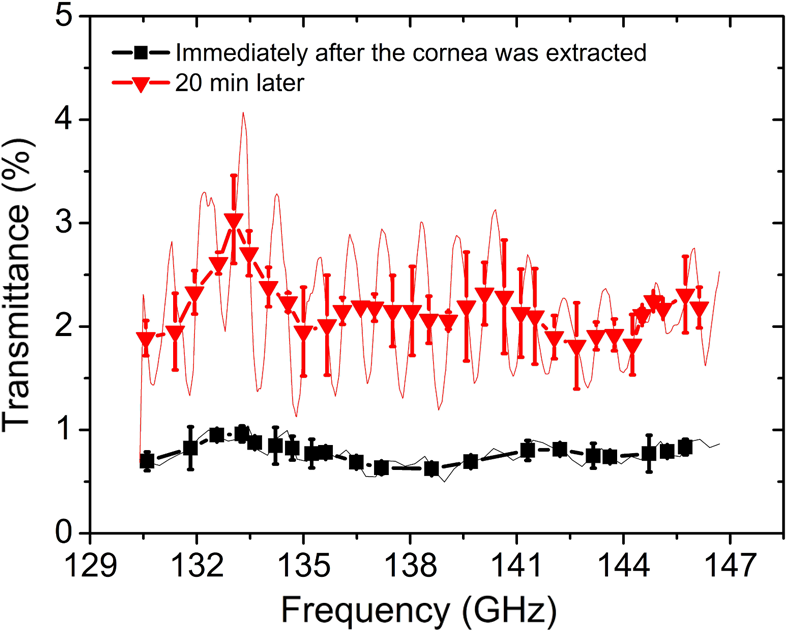 Fig. 6The transmittance spectrum of rabbit sclera. The black curve shows the values immediately after the sclera was extracted from the eye, while the red curve was obtained 1 h later.  Figure 9 shows the results of the experiments measuring the reflectance dependence of the rabbit cornea and sclera on the content of free water in the tissue. The free water content in the rabbit cornea and sclera was obtained using the measured mass of the tissue at every particular point on the plot and the measured mass of the completely dry tissue according to the equation . The measurements were carried out for more than 1.5 h. We obtained 35 data points for each experimental curve. A part of the radiation reflected from the material could not be detected due to nonideal coupling with the detector and scattering. For this reason, the reflected signal is expressed in relative units in Fig. 9. Fig. 9The curve showing how the reflectivity depends on the water content in the tissue. (a) The reflected signal drops with the decrease of free water concentration in the tissue. (b) The linear part of the dependence. 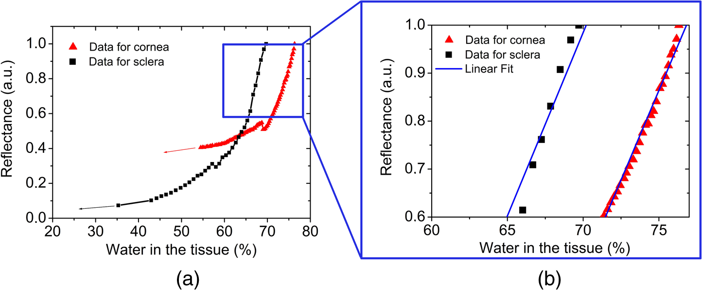 As can be seen from Fig. 9, the decrease of free water concentration by 1% leads to a clearly detectable drop of the reflected signal by 13%. This result agrees well with that obtained earlier in an experiment with pulsed THz system.18 The linear dependence of the reflectivity on the mass of water content in tissue is also confirmed at the initial stages of drying. A stronger drying of cornea and sclera leads to a change in the shape of the curve, which is probably connected with the deformation of the cornea itself, caused by dehydration. The obtained result is promising in terms of the possibility of developing a practical device for noninvasive analysis and control of the hydration degree of the cornea and the sclera. 4.ConclusionThe presented work reports on the creation, for the first time, of experimental installations for in vitro studies of the frequency dependence of the transmittance coefficient of the cornea and the sclera, as well as the dependence of reflectance coefficient on the content of water mass in these tissues in the sub-THz frequency range. The reflection spectrum of the eye was measured in the 0.13 to 0.32 THz. The transmittance spectra of the cornea and the sclera and the dependence of the reflection coefficient of these tissues on water content were obtained. Differences have been found in the investigated parameters between the cornea and the sclera of the rabbit, as well as those between the rabbit sclera and the human sclera. The preliminary results show that the proposed technique, based on the use of continuous THz radiation, can be used to create a setup for noninvasive control of the degree of hydration of the cornea and the sclera, which has a clear potential of broad practical implementation in clinical ophthalmology. AcknowledgmentsThis work was supported in part by the Russian Foundation for Basic Research (RFBR) (Grant Nos. 15-29-03874 and 16-32-00622) and by the Ministry of Education and Science of the Russian Federation (Grant No. 14.B25.31.0007). Professor Dr. Iomdina has nothing to disclose. ReferencesV. V. Serov and A. B. Shekhter, Connective Tissue: Functional Morphology and General Pathology, Meditsina, Moscow
(1981). Google Scholar
H. Fessler,
“Water and mucopolysaccharide as structural components of connective tissue,”
Nature, 179 426
–427 1957). http://dx.doi.org/10.1038/179426b0 Google Scholar
I. Fatt and B. A. Weissman, Physiology of the Eye: An Introduction to the Vegetative Functions, Butterworth-Heinemann, Waltham, Massachusetts
(2013). Google Scholar
C. Costagliola et al.,
“Corneal oedema and its medical treatment,”
Clin. Exp. Optometry, 96
(6), 529
–535 2013). http://dx.doi.org/10.1111/cxo.12060 Google Scholar
G. C. Lim et al.,
“Toxic keratopathy-related corneal dehydration after laser in situ keratomileusis,”
J. Cataract Refractive Surg., 31
(8), 1656
–1658
(2005). http://dx.doi.org/10.1016/j.jcrs.2005.01.020 Google Scholar
E. S. Avetisov et al.,
“A study of biochemical and biomechanical qualities of normal and myopic eye sclera in humans of different age groups,”
Metab., Pediatr. Syst. Ophthalmol., 7
(4), 183
–188
(1983). MPSODY 0277-9382 Google Scholar
N. A. McBrien and A. Gentle,
“Role of the sclera in the development and pathological complications of myopia,”
Prog. Retinal Eye Res., 22
(3), 307
–338
(2003). http://dx.doi.org/10.1016/S1350-9462(02)00063-0 Google Scholar
J. Rada, S. Shelton and T. T. Norton,
“The sclera and myopia,”
Exp. Eye Res., 82
(2), 185
–200
(2006). http://dx.doi.org/10.1016/j.exer.2005.08.009 EXERA6 0014-4835 Google Scholar
E. N. Iomdina, N. Y. Ignatieva and N. A. Danilov,
“Biochemical and structural peculiarities of human sclera matrix in primary open angle glaucoma,”
Vestnik oftal’mologii, 6 10
–14
(2011). Google Scholar
N. A. Danilov et al.,
“Sclera of the glaucomatous eye: physicochemical analysis,”
Biofizika, 56
(3), 520
–526
(2011). http://dx.doi.org/10.1134/S0006350911030067 BIOFAI 0006-3029 Google Scholar
B. Coudrillier et al.,
“Biomechanics of the human posterior sclera: age- and glaucoma-related changes measured using inflation testing,”
Invest. Ophthalmol. Visual Sci., 53
(4), 1714
–1728
(2012). http://dx.doi.org/10.1167/iovs.11-8009 IOVSDA 0146-0404 Google Scholar
P. Tewari et al.,
“Terahertz sensing of corneal hydration,”
in Annual Int. Conf. IEEE on Engineering in Medical and Biology Society (EMBC 2010),
3021
–3024
(2010). http://dx.doi.org/10.1109/IEMBS.2010.5626146 Google Scholar
S. Seliverstov et al.,
“Fast and sensitive terahertz direct detector based on superconducting antenna-coupled hot electron bolometer,”
IEEE Trans. Appl. Supercond., 25
(3), 1
–4
(2015). http://dx.doi.org/10.1109/TASC.2014.2372171 ITASE9 1051-8223 Google Scholar
Z. D. Taylor et al.,
“THz imaging based on water-concentration contrast,”
Proc. SPIE, 6949 69490D
(2008). http://dx.doi.org/10.1117/12.785337 PSISDG 0277-786X Google Scholar
V. P. Wallace et al.,
“Terahertz pulsed imaging of basal cell carcinoma ex vivo and in vivo,”
Br. J. Dermatol., 151
(2), 424
–432
(2004). http://dx.doi.org/10.1111/j.1365-2133.2004.06129.x BJDEAZ 0007-0963 Google Scholar
J. P. Dougherty, G. D. Jubic and Jr. W. L. Kiser,
“Terahertz imaging of burned tissue,”
Proc. SPIE, 6472 64720N
(2007). http://dx.doi.org/10.1117/12.705137 PSISDG 0277-786X Google Scholar
D. Mittleman, Sensing with Terahertz Radiation, 85 Springer, New York
(2013). Google Scholar
D. B. Bennett et al.,
“Terahertz sensing in corneal tissues,”
J. Biomed. Opt., 16
(5), 057003
(2011). http://dx.doi.org/10.1117/1.3575168 JBOPFO 1083-3668 Google Scholar
Z. D. Taylor et al.,
“THz and mm-wave sensing of corneal tissue water content: in vivo sensing and imaging results,”
IEEE Trans. Terahertz Sci. Technol., 5
(2), 184
–196
(2015). http://dx.doi.org/10.1109/TTHZ.2015.2392628 Google Scholar
BiographyElena N. Iomdina is the principal researcher in Moscow Helmholtz Research Institute for Eye Diseases. Her main research interests include myopia pathogenesis, surgical and nonsurgical methods of myopia treatment and sclera reinforcement in progressive myopia, research in glaucoma pathogenesis and treatment, biomechanics and biochemistry of the eye (particularly the cornea and the sclera), and experimental ophthalmology. She has authored over 150 articles in peer reviewed journals, a book on eye biomechanics, and 40 patents and inventions. Gregory N. Goltsman is a professor and a leading researcher in the Physics Department of Moscow State Pedagogical University (MSPU). He received his PhD in 1973 and his Dr. habil. in 1985. His scientific interests include superconductivity, applied physics, and terahertz technology. He has extensive experience in industry, where he has contributed to the development of small innovative hi-tech businesses. He is the author and coauthor of more than 200 publications. Sergey V. Seliverstov is a researcher in the Physics Department of MSPU. His scientific interests include a nonequilibrium superconductivity, terahertz radiation, and applied physics. The focus of his work is on the interaction of terahertz radiation with nanoscale objects, primarily superconductors. Alisa A. Sianosyan is a PhD student at Moscow Helmholtz Research Institute for Eye Diseases. Her main scientific interests include myopia pathogenesis, biomechanics, and biochemistry of the eye. |


