
|
|
1.IntroductionRoot canal infection, also known as apical periodontitis involves inflammation and destruction of periradicular tissues adjacent to the root apex of a tooth. This is primarily caused by the presence of bacterial biofilms within the root canal system of the tooth.1, 2 Apical periodontitis is not self-healing, since microbes established in the “secluded” sanctuaries of the root canal system cannot be completely resolved by the host immune defense.3 The principal treatment involves the elimination of microbial flora from the root canal system. Conventionally, disinfection of the root canal is achieved by combining mechanical instrumentation with caustic chemicals4 (the chemomechanical approach). Nevertheless, complete disinfection of the root canal is not achieved, though in most cases the clinical symptoms recede.5, 6 Major factors limiting the elimination of bacteria by conventional treatment are the inability of chemical disinfectant to destroy bacteria residing in the dentinal tubules and anatomical complexities of the root canal, and the inability of the antimicrobial agents to eliminate certain species of bacteria and bacterial biofilm.6, 7, 8 The biochemical composition of biofilm matrix and the altered physiology of biofilm bacteria may contribute toward the observed resistance to antimicrobial agents.8, 9, 10 In fact, Enterococcus faecalis has shown the ability to produce biofilm on root canal wall medicated with calcium hydroxide, one of the most widely used intracanal medicament.11 In addition to the preceding shortcomings, the indiscriminate use of caustic chemicals has been reported to adversely affect the chemical and mechanical properties of dentine.12, 13 Recently, light-activated therapy (LAT), generally known as photodynamic therapy, is showing great potential in the treatment of localized bacterial infections.14, 15 The killing of bacteria by LAT can be induced by two photoreactions: (1) a type I reaction, where the electron transfer between the triplet state sensitizer and biomolecules results in the generation of several radical species, which can cause cell damage, and (2) a type II reaction, where the energy transfer from the triplet state sensitizer to molecular oxygen produces singlet oxygen , a highly reactive species causing cell death.16 The activation of photosensitizers in the presence of oxygen results in the production of reactive oxygen species, involving hydroxyl radicals, superoxides, and singlet oxygen.15, 17 The oxygen-based free radicals involved in the photooxidized inactivation of microorganisms acts on multiple target in a bacterial cells, resulting in instantaneous killing.18 This aspect of LAT makes the selection of resistant microbial strain highly unlikely.16, 17 Production of highly reactive singlet oxygen capable of destroying biomolecules has been identified as the principle agent causing bacterial killing. LAT has the ability to destroy a wide range of microbial pathogens that includes wild and antibiotic resistant gram positive bacteria, gram negative bacteria, candida, viruses, and protozoa. 19, 20, 21, 22, 23 It is, therefore, suggested as an alternative to conventional antimicrobial agents in combating polymicrobial infections involving biofilms. 15, 17, 18, 24 Many studies have shown the successful application of LAT in the elimination of bacteria associated with periodontal diseases.18, 25, 26 Recently, a few studies were also carried out to achieve root canal disinfection.27, 28 These studies highlighted the advantages of using LAT to kill residual bacteria in the root canal system after conventional root canal chemomechanical treatment. However, as seen from this studies, when used alone, LAT could not produce complete reduction in bacterial population. The efficiency of LAT may depend on the environmental and microbiological factors at the site of infection. It is known that an infected root canal harbors a polymicrobial population involving aerobic, anaerobic, gram positive, and gram negative bacteria.29 The susceptibility of bacteria to LAT depends on the type of cell wall they posses. Gram positive bacteria are reported to be more susceptible to LAT-based killing compared to gram negative bacteria, which possess an outer membrane.16, 30 Another important aspect of root canal infection is the dynamic nature of microbial flora along with shift in the environmental conditions.2 As the infection advances, the oxygen tension in the root canal reduces and microbial flora shift to more of anaerobic type.2, 29 The reduced oxygen tension in the root canal may lower the efficiency of LAT since the singlet oxygen generated from the molecular oxygen is the principle antimicrobial agent. In addition to the microbiological and environmental factors, other factors that limit LAT in root canal disinfection are (1) the anatomical and geometric complexities of the root canal, (2) the porous nature of the dentine, and (3) interactions of photosensitizer molecules with each other and other molecular entities at the target site.31, 32 All the preceding tissue-specific factors necessitate the use of an ideal photosensitizer formulation and a treatment strategy for root canal disinfection. This study aimed to investigate the photophysical, photochemical, and photobiological properties of methylene blue (MB) in different formulations for root canal disinfection. MB was chosen as the photosensitizer since its activity against oral bacteria is known and has reported clinical application.33 The absorbance characteristics and aggregation of MB was studied to assess the effect of different formulations on the photophysical behavior of MB. Photochemical characteristics such as model substrate oxidation and singlet oxygen production were studied to determine the photooxidation performance of MB in different formulations. Finally, a novel dual-stage approach of using a photosensitizing medium and an irradiating medium to enhance light-activated disinfection of bacterial biofilms in the root canal was evaluated. 2.Materials and MethodsAll the chemicals used in the study were of analytical grade and were purchased from Sigma Aldrich, St. Louis, Misouri, USA, unless noted. MB a phenothiazine dye used as the photosensitizer was tested in four different formulations, namely, water (deionized water), 70% glycerol in water (glycerol), 70% poly ethylene glycol-200 in water (PEG), and a mixture of glycerol:ethanol:water (30:20:50) (MIX). The multicomponent formulation of MIX is expected to facilitate the photosensitizer diffusion into dentinal tubules and anatomical complexities of root canal. Glycerol and ethanol are widely used vehicles for various commercial products such as drugs, cosmetics, and foods. Pilot experiments showed the tested ratio of glycerol:ethanol:water to be optimum for photosensitizer uptake by bacterial cells and bactericidal action on irradiation. A diode laser of wavelength with an output energy of was used as the light source. The wavelength of the light source corresponded with the excitation wavelength of MB. The laser light was delivered using an optical fiber of m outer diameter (LDCU/6130, Power Technology Inc, Little Rock, Arkausas, USA). 2.1.Photophysical Characteristics of MB in Different FormulationsIn this experiment, the absorption spectra of 100- M MB in different formulations were determined using a UV/VIS spectrophotometer (Shimadzu 1100, Japan, cuvette pathlength ). The ratio of absorbance at 664 nm to absorbance at was calculated and plotted as an index of monomer to dimer in different formulations at increasing concentrations34 of 1, 5, 15, 20, 25, 50, and M. The mean ratio was calculated from three independent absorbance readings. 2.2.Photochemical Reactions of MB in Different Formulations2.2.1.Model substrate oxidationThe photosensitizing activity of MB (in different formulations) was evaluated fluorimetrically using35 the photooxidation of model substrate -acetyl-L-tryptophanamide (NATA). NATA is a widely accepted model molecule to test the efficiency of photosensitizing agents. Oxidation of NATA can be caused by type 1 or type 2 mechanisms of photosensitization. MB in tested formulations containing NATA was taken in a fluorimetric cuvette and the fiber tip was kept as close as possible to the liquid surface without touching. Irradiation was carried out with a diode laser with a power of . The test solution was maintained undisturbed during the experiment. The rate of decrease of NATA concentration with increase in duration of irradiation (5-min intervals for ) was monitored by measuring the intensity of -excited fluorescence emission spectrum (300 to ) typical of the tryptophanyl moiety. 2.2.2.Singlet oxygen measurementSinglet oxygen measurements were carried out in a quartz cuvette according to a procedure described previously.36 The assessment of different formulations of MB generating singlet oxygen on photoactivation was studied photometrically using 1,3-diphenylisobenzofuran (DPBF), a singlet oxygen scavenger.36, 37 DPBF at a concentration of M, corresponding to absorbance intensity between 1.5 and 2 at , was mixed with M MB in different tested formulations (total volume, ). The assay was performed as in the preceding experiment without dipping the fiber tip. The decrease in absorbance intensity at was monitored for an irradiation period of (at 5-min interval) using a UV-VISIBLE spectrophotometer (Shimadzu, Japan). The rate of singlet oxygen production was related to the rate of decrease of DPBF absorbance at as a function of irradiation time. 2.3.Photobiological Properties of MB in Different Formulations2.3.1.Penetration of MB into dentinal tubulesThe institutional review board of the National University of Singapore approved the collection and use of extracted human teeth for all the experiments conducted in this study. Twelve tooth specimens with no history of caries and/or defects were selected. The crown portion of all samples was sectioned at the level of cementoenamel junction, to obtain an approximate length of . Patency of the root canal was obtained by widening the root canal minimally using endodontic files (K files #20 to #40). The smear layer that formed on the root canal wall during mechanical preparation was removed by alternate washing with 1% sodium hypochlorite and ethylene diamine tetra acetic acid (EDTA) (Merck, USA). These specimens were washed and kept upright in 96 well plates containing semisolidified 2% agar. Solidified agar stabilized the tooth in the socket formed. MB at a concentration of , in different media, was introduced into the root canals by using a syringe and was kept at 37°C for . The excess MB remaining in the root canal was blotted using paper points. Thin sections (cross sections) of these specimens were prepared by using a microslice machine (Metal Research, England). These cross sections were scanned using HP Scanjet 3970 (Hewlett-Packard, California, USA). The extent of diffusion of MB was measured using UTHSCSA Image Tool (version 3) software. MB diffusions into dentinal tubules were measured at three regions along the root canal length, namely, cervical (first three sections of a single root), middle (three sections from middle portion of the specimen), and apical (last three sections of a single root). Three regions of interest were used in this analysis since penetration of liquid into the dentinal tubules is influenced by the taper of the root canal and diameter of dentinal tubules, which varies from the cervical to the apical region. The extent of MB penetration into dentinal tubules was expressed as the percentage distance diffused across the entire length of dentinal tubules. This was calculated by dividing the total distance diffused by MB from the lumen of the root canal by the total length of the dentine bulk from the lumen to the outer surface. A relatively high concentration of MB was used to facilitate the visual examination of the photosensitizer penetration into the dentinal tubules. 2.3.2.Uptake of MB by bacterial cellsThe MB uptake by bacterial cells was conducted as described elsewhere with certain modifications.38 Enterococcus faecalis (ATCC 29212), a gram positive bacteria, and Actinobacillus actinomycetemcomitans (ATCC 33384), a gram negative bacteria, were used in this study. Bacterial cultures were grown aerobically in brain heart infusion broth (BHI). Cells in the stationary phase of growth were harvested by centrifugation ( for ) and were washed with deionized water. The optical density of the culture was adjusted to 1 at that corresponded to . Cells harvested from of the preceding suspensions were mixed with of M MB in different test formulations and were incubated at 37°C for in an orbital incubator . These cells were harvested, washed twice with deionized water, as already described, and lysed by treating with of 2% sodium dodecyl sulfate (SDS) for . The absorbance intensity of the supernatant solution after centrifugation ( for ) was recorded at .The percentage of MB taken up by bacterial cells from the original solution was calculated from the calibration curve plotted with known concentrations of MB in SDS. 2.4.LAT on Biofilm Bacteria2.4.1.LAT on bacterial biofilms grown in multiwell platesTwo-day-old biofilms of E. faecalis and A. actinomycetemcomitans were produced in wells of multiwell plates (material-polystyrene) using all culture (AC) and BHI media, respectively, as nutrient sources. After the incubation period, the growth media were removed and wells were rinsed with deionized water, retaining the biofilm bacteria in the well. Biofilm growth was evident on the walls of the wells on visual examination. The entire process of LAT was conducted in two steps. In the first step, the biofilm bacteria formed on the wall of multiwell plate well was sensitized with M MB in test formulations (photosensitizing media) for . The volume of photosensitizing media was same as the original volume of growth media ( L) to cover the biofilm produced in the well. Subsequently, the excess photosensitizer media was removed using a micropipette, leaving behind the biofilm-bound MB. In the second step, wells were added with L of oxygen carrier solution of perfluro-decahydro naphthalene (irradiation medium), and were irradiated for with the same light source used in previous experiments. During irradiation, the tip of the optical fiber was placed just above the irradiating media and the plate was shaken using a plate shaker. The total energy delivered from the tip of the optical fiber was . After treatment, the liquid in the wells were replaced with fresh growth media and vigorously shaken. Growth media was flushed using a micropipette for mechanically disrupting the biofilm bacteria on the walls of the wells. Serial dilutions were carried out in the growth medium, and L from each dilution was spread plated on to AC agar to enumerate the surviving bacterial cells. Control wells were added with phosphate-buffered saline (PBS) instead of either of the test media and were not subjected to light activation. Wells treated with either light alone or photosensitizer alone were also included in the study to assess their bactericidal effect. In the second step of LAT, an irradiation medium was used to create a liquid conduit within the root canal lumen and dentinal tubules, when applied in teeth. The liquid conduit intends to minimize attenuation of light energy and enable greater penetration of light into the dentine. The materials for the liquid conduit is chosen in such a way that they are transparent, have a low refractive index, are biologically inert, and can be a source of dissolved oxygen. In this study, we used perfluro-decahydro naphthalene, which satisfies the preceding requirements.39, 40 Perfluro-decahydro naphthalene in addition to its oxygen carrier effect will also increase the life time of singlet oxygen.41 These factors will further enhance the efficacy of the photodynamic effect. 2.4.2.LAT on bacterial biofilms grown in tooth specimensThe methodology of the LAT experiment conducted in a root canal is summarized in Fig. 1 . Seventy-two single-rooted noncarious teeth were collected. They were prepared by removing the crown at the level of cemento-enamel junction, and the apical third of the root to obtain a standard length of . To minimize variation in the internal dimensions of root canals and for standardization, all specimens were instrumented using K files with sizes from #20 to #40. The smear layer formed during mechanical shaping was removed by rinsing of root canal with sodium hypochlorite (1%) followed by EDTA . After thorough washing with deionized water, these tooth specimens were randomly divided into two equal groups. Specimens in one group were incubated with of AC media inoculated with a single colony of E. faecalis and those in the second group were incubated with of BHI media inoculated with a single colony of A. actinomycetemcomitans. Incubation at 37°C under constant shaking for 4 days since biofilm formation in dentine takes more time than in a multiwell plate. The growth medium was replaced with fresh on every 2 days. Early studies from our lab have shown that biofilm can be formed on root canal wall during this time interval. For the LAT experiment, tooth specimens with bacterial biofilm in the root canal were removed from the culture medium and the surface was wiped with 70% ethanol. These specimens were positioned with the coronal root canal orifice facing superiorly by embedding them in 2% agar in wells of 24 wellplates. This positioning of tooth specimens was similar to clinical situation, where the root portion of a tooth is housed within the alveolar bone socket. At the outset, the culture medium remaining in the root canal was removed by using adsorbent paper points. LAT was performed in two steps, as with the in vitro experiment conducted previously. Photosensitization was carried out for at 37°C by keeping the photosensitizing media ( MB) in the root canal. The chosen photosensitization time in this experiment ensured penetration of MB into dentinal tubules of tooth. Subsequently, the photosentizing media was removed from the root canal using adsorbent paper points, and the root canal space was filled with the irradiation media. Irradiation was carried out with a diode laser of wavelength coupled to an optical fiber, as in previous experiments. The optical fiber terminus was placed at the coronal end of the root canal and irradiation was carried out for (total energy, ). Later, the irradiation media was removed, and grooves were prepared on the proximal sides of the tooth specimens. Tooth specimens were split opened using a chisel and mallet. A round bur of diameter, held perpendicular to the root canal surface, was used to obtain dentine shavings harboring the bacteria.42 Dentine shavings were collected up to the working length of the bur to obtain bacteria in root canal wall and those in dentinal tubules. These shavings were obtained from the root canal wall apical to the root canal orifice (Fig. 1). The dentine shavings were added to fresh AC/BHI media and mixed by vortexing. This was incubated at 37°C for to enrich the number of bacteria obtained from the dentine shavings. Serial dilutions were prepared in growth medium and L from each dilution was spread plated on to AC agar. The bacterial counting was carried out after of incubation at 37°C. The control specimens received no light or photosensitizer treatment. One set of tooth specimens were subjected to irradiation alone with the root canal lumen filled with PBS during irradiation. 3.Results3.1.Photophysical Characteristics of MB in Different FormulationsFigure 2 shows the absorption spectra of MB in different formulations. The spectrum showed two bands peaking at 612 and corresponding to the dimer and monomer, respectively. The peak corresponding to the dimer was most prominent when MB was dissolved in water. As the molar concentration of MB was increased from 1 to M, the absorption characteristics of MB changed. The peak corresponding to the dimers became more prominent along with the concentration, indicating the aggregation of photosensitizer molecules (Fig. 3 ). MB dissolved in water showed a linear reduction in the monomer-to-dimer ratio. The monomer-to-dimer ratio was significantly lower in water concentrations above M compared to all other formulations. At a M concentration there was no significant difference in the ratio among glycerol, PEG, and MIX-based formulations. 3.2.Photochemical Reactions of MB in Different Formulations3.2.1.Model substrate oxidationThe rate of NATA oxidation caused by photoactivation of MB in different formulations is given in Fig. 4 . Under the experimental conditions, the NATA oxidation caused by photoactivation of MB in MIX was found to follow First-order kinetics. The rate of NATA oxidation when MB was dissolved in MIX was significantly higher than any other formulation . MB dissolved in water also showed significant NATA oxidation. There was no significant decrease in NATA concentration when MB was dissolved in PEG and glycerol. 3.2.2.Singlet oxygen yieldThe rate of DPBF bleaching is shown in Fig. 5 , representative of the rates of singlet oxygen production. The raw kinetic data was in the form of concentration of DPBF (from absorbance value at ) as a function of time in minutes. The ability to produce DPBF bleaching species (singlet oxygen) in different formulations was in the order MIX glycerol water PEG. The MIX-based formulation showed threefold increased rate of DPBF bleaching compared to other formulations, indicating a higher rate of singlet oxygen production. Fig. 5Decreases of DPBF concentration when MB dissolved in different formulations was irradiated for increasing time intervals. The rate of decrease of DPBF concentration is correlated to the rate of singlet oxygen production. The value for oxidation of DPBF was significantly higher with MB dissolved in MIX compared to other formulations .  3.3.Photobiological Properties of MB in Different Formulations3.3.1.Penetration of MB into dentinal tubulesThe percentage of MB penetration across the entire length of dentinal tubules is shown in Fig. 6 . The penetration of photosensitizer formulations into the dentinal tubules were examined from three regions of interest, namely, the coronal, middle, and apical portions of the root canal (Fig. 7 ). MB in MIX formulation showed the maximum penetration in all the regions of interest under the test condition, which was significantly higher than MB in water . MB dissolved in water showed the least penetration across the dentinal tubules. Fig. 6Penetration of MB into dentinal tubules when applied in different formulations. Three different regions along the length of root canal were analyzed for MB penetration. The formulation based on MIX showed the maximum diffusion across the dentinal tubules in all the three regions analyzed. The error bars show standard deviation from average value.  3.3.2.Uptake of MB by bacterial cellsThe uptake of photosensitizer by bacterial cells, expressed as percentage MB uptake, is shown in Fig. 8 . Here the absorbance at corresponding to original concentration of MB applied ( M) was taken as 100%. The photosensitizer uptake was significantly higher in water compared to all other formulations , which was followed by MIX. All the formulations showed an increased photosensitizer uptake by E. faecalis compared to A. actinomycetemcomitans, although it was not statistically significant in water alone. Fig. 8Percentage of MB taken up from M of original MB formulation by bacterial cells ( to ). There was significant variation in uptake of photosensitizer by bacterial cells when applied in different formulations. We found E. faecalis to have higher MB uptake (gram positive bacteria) compared to A. actinomycetemcomitans (gram negative) . Error bars show the standard deviation from average value. 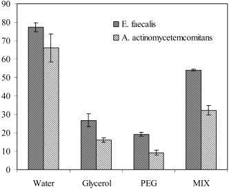 3.4.LAT on Bacterial Biofilms3.4.1.LAT on biofilms grown in multiwell platesFigure 9 shows the number of E. faecalis surviving after the LAT treatment. Under the experimental conditions, 100% killing of E. faecalis and A. actinomycetemcomitans was obtained when LAT was conducted using MIX based MB formulation. Further, it was the combination of photosensitizer and irradiation that produced the desired effect of bacterial killing. Neither laser treatment nor photosensitizer formulations alone had any significant effect on bacterial killing. Fig. 9Log number of E. faecalis and A. actinomycetemcomitans cells in biofilm surviving the LAT conducted in multiwell plate. The LAT with MB in MIX showed complete killing of both bacteria on irradiation. Compared to the control group, the reduction in the number of E. faecalis cells was significant for all treatment group except for the glycerol-based formulation, and that of A. actinomycetemcomitans was significant only in MIX-based formulation . There was no significant reduction in the number of bacterial cells when formulation alone was used. Error bars show the standard deviation from average value.  3.4.2.LAT on bacterial biofilms grown in tooth specimensThe result of LAT on biofilms developed on root canals showed varying percentages of bacterial killing according to the formulation used (Fig. 10 ). The pattern of bacterial killing by formulations was similar to those seen in biofilm developed in the multiwell plate. Unlike in the multiwell plate, the exposure of tooth specimens to light alone also resulted in significant reduction in colony-forming units (CFUs). In the case of E. faecalis, the formulation based on MIX was found to produce 100% bacterial killing, as revealed by the CFU counting. Significant bacterial reduction was also observed in formulation based on PEG. LAT conducted in root canals with A. actinomycetemcomitans biofilms showed similar trends. The formulation based on MIX showed 99.77% reduction in bacterial count compared to the control group. In general, the susceptibility of A. actinomycetemcomitans to the bactericidal action of LAT was significantly less compared to that of E. faecalis. Fig. 10Log number of E. faecalis and A. actinomycetemcomitans cells in biofilm in root canal dentine, surviving LAT. The reduction in the number of E. faecalis cells was significant for all treatment groups except for glycerol-based formulation and that of A. actinomycetemcomitans was significant for only MIX, water, and glycerol . Error bars show the standard deviation from average value. 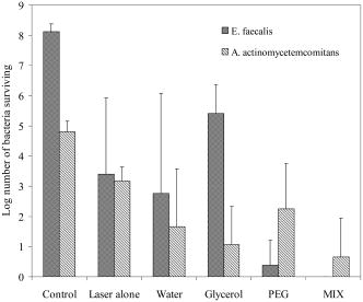 4.DiscussionAlthough many studies have shown the lethal effect of LAT on oral microorganisms, only a few addressed the feasibility of applying this technique for root canal disinfection. The predominance of anaerobic bacteria in advanced endodontic infection make a LAT-based regimen that produces oxygen free radicals a promising treatment strategy. The difficulty in achieving complete disinfection of a root canal by light activation in the earlier investigations highlights the fact that the mere combination of photosensitizer with specific wavelength of light is incomplete by itself.27, 43 In this regard, tissue-specific modifications of LAT was suggested to achieve maximum killing of bacteria. This study was conducted with the intention of enhancing the light inactivation of biofilm bacteria by MB for its possible application in root canal disinfection. An ideal formulation and/or strategy that can ensure proper delivery of the photosensitizer, enhance photophysical characteristics, and stabilize oxygen free radicals would account for a better outcome. Results from our study showed that the light absorption property of MB varied in different formulations. When dissolved in water, the absorption spectra of MB at a M concentration displayed peak maxima corresponding to the dimer. The data suggest that the polarity of the formulation affected the absorbance characteristics of MB. Formulations based on PEG, glycerol, and MIX showed a tendency to prevent photosensitizer aggregation, as is evident from the monomer-to-dimer ratio. Earlier studies conducted by Severino have shown that the photophysical and photochemical characteristics of MB were modulated by the amount and the type of dimer formed.44 Since dimer formation may decrease the production of singlet oxygen, a formulation that stabilizes monomer is preferred for therapeutic purpose.34, 45 Further, the membrane potential of cells may also influence the monomer-to-dimer ratio of MB and its photochemical behavior, as seen with mitochondrial suspension.34 The photooxidation potential of MB was studied in different formulations. The photochemical properties of MB clearly indicated that the behavior of the electronically excited state of MB was highly influenced by the formulation used. When dissolved in MIX, the oxidation of NATA and DPBF showed first-order kinetics, typical of the photooxidation reaction. The increased photooxidation potential of MB in MIX could be due to the presence of an apolar solvent such as ethanol. The half-life of singlet oxygen is considerably longer in ethanol ( s) compared to that in water18 ( s). The oxidation of NATA and DPBF was found to be significantly less in PEG and glycerol-based formulations, though the monomer concentration was relatively high. This variation could be due to the interaction of the excited state of photosensitizer with the solvent molecules. However, further study is indicated toward understanding the type and extent of solvent effect on the excited state of photosensitizer molecule. The photochemical behavior of MB in water showed a relatively high rate constant for NATA oxidation compared to that of DPBF. Since NATA can be oxidized by radicals others than singlet oxygen but not DPBF, the most plausible explanation could be the favoring of a type 1 photoreaction under this condition. Additional support for this statement derives from the observation that MB aggregation leads to the production of radical formation instead of singlet oxygen.45 Thus, the relatively high proportion of aggregated MB in water may favor formation of radicals instead of singlet oxygen. The inability to eliminate a bacterial population from anatomical complexities and dentinal tubules is one of the serious shortcomings in current root canal disinfection. Therefore, adequate diffusion of photosensitizer into anatomical complexities and dentinal tubules is a prerequisite for endodontic disinfection. Results from the experiments conducted to study the diffusion of MB into dentinal tubules showed that the formulation based on MIX facilitated maximum penetration into dentinal tubules. A penetration depth greater than in the coronal region was seen with MB dissolved in MIX. Earlier studies have indicated that the maximum depth of bacterial penetration into the dentinal tubules in this area seldom goes beyond , even after growing bacteria under in vitro conditions for 4 weeks.46 Hence, by applying a MIX-based formulation, it is possible to target bacterial cells invading the dentinal tubules. Enhancing the uptake of photosensitizer by the bacterial cells is another important factor influencing the effectiveness of LAT-based bacterial killing. The limited diffusion distance and half-life of singlet oxygen necessitate the close proximity of the target molecule.15 An ideal formulation should facilitate the photosensitizer uptake by both gram negative and gram positive bacteria as the infected root canal may harbor a multibacterial consortium. The presence of an outer membrane in gram negative bacteria prevent drug or photosensitizer uptake and they are relatively less susceptible to light-activated killing.16, 20 The uptake of MB was significantly less in A. actinomycetemcomitans cells compared to that of E. faecalis. The water-based MB formulation showed relatively higher uptake of MB by both organisms. However, the localization of MB in bacterial cells when applied in different formulation could be different. Finally, the effectiveness of different photosensitizer formulations in combination with irradiation media was tested for their bactericidal action on biofilm grown under in vitro and ex vivo conditions. Among the different formulations tested, the MIX-based formulation showed a better photodynamic effect, as is evident from the bacterial killing. The replacement of photosensitization media with irradiation media during the irradiation stage was not expected to alter the photophysical and photochemical behavior of MB due to the immiscible nature of irradiation media with photosensitization media. Hence, the local concentration and the photosensitizer’s behavior in the formulation could be the reason for the variation in bacterial killing observed between different groups. If the formulation promotes MB binding to the outer surface of bacteria, it could result in a localized increase in the photosensitizer concentration, as suggested by Gabrielli .34 Subsequently, the aggregated MB would lower the quantum yield of singlet oxygen, resulting in decreased bacterial killing. This could be the possible reason for the decreased bacterial killing observed with MB in water, although the MB association was more with bacterial cells in the water-based formulation. Furthermore, light alone produced no effect on viability of biofilm bacteria in a multiwell plate, however, there was a considerable reduction in CFU when irradiation was conducted in tooth specimens. Unlike the transparent material of the multiwell plate, the optical characteristics of dentine, such as light absorption and scattering, could be the apparent reasons for this observation. However, further experiments are recommended to ascertain this observation. Data from our study showed that it is possible to achieve complete killing of E. faecalis cells embedded in biofilm formed on animate and inanimate material. Enterococcus faecalis is recognized as one of the human pathogens with a high resistance to many antimicrobial agents. Although the presence of E. faecalis in primary endodontic infection is comparatively less, it has been isolated in high proportion or as single isolate in therapy failed root canals.47, 48 The findings from our study showed that the LAT of A. actinomycetemcomitans biofilm was less effective compared to E. faecalis biofilm, although MIX showed 99.77% killing. The lower effectiveness of LAT in eliminating A. actinomycetemcomitans may be due reduced MB uptake, as seen in earlier experiments. Increasing the light energy, and duration of photosensitization and the use of a more potent photosensitizer are possible alternatives to enhance the efficacy of LAT on gram negative bacteria. Nevertheless, the presented experiment showed better treatment outcome than other reported photodynamic-based killing of biofilm bacteria.25, 49 The considerable bacterial killing in root canal dentine observed in this study can be attributed to the photosensitizer formulation and the application of photosensitizing and irradiation media in two stages. The application of photosensitizing media in the first stage facilitated enhanced penetration of the photosensitizer into the dentinal tubules and anatomical complexities, while ensuring ideal uptake of the photosensitizer by the bacterial cells. The irradiation medium used in the second stage created a liquid conduit within the root canal lumen and the dentinal tubules. This medium minimized attenuation of light energy and enabled greater penetration of light into the dentinal tubules. Since the improved LAT based on an MIX formulation showed maximum killing of bacteria in biofilm produced on an inanimate substrate and root canal dentine, its application as a root canal treatment regimen is highly recommended. In addition this investigation highlights the importance of tissue-specific characterization of LAT components for better treatment outcome. In conclusion, although MB uptake by bacterial cells was high when applied in a water-based formulation, the photochemical assays showed that MIX-based formulation had a better photooxidation potential. The MB diffusion into dentinal tubules also revealed the advantages of MIX-based formulation. The disinfection of bacteria in a biofilm produced on tooth specimens and inanimate material showed a formulation-depended effect, as predicted from the photophysical and photochemical studies. The dual-stage application of an appropriate photosensitization medium and irradiation medium aided in achieving more thorough disinfection. AcknowledgmentFunding from the National University of Singapore (ARF), Grant No. FY 04 R-224-000-021-101, is gratefully acknowledged. ReferencesP. N. R. Nair,
“Light and electron microscopic studies of root canal flora and periapical lesions,”
J. Endod., 13 29
–39
(1987). 0099-2399 Google Scholar
L. Tronstad and
P. T. Sunde,
“The evolving new understanding of endodontic infections,”
Endodont. Top., 6 57
–77
(2003). Google Scholar
P. N. R. Nair,
“Pathogenesis of apical periodontitis and the cause of endodontic failures,”
Crit. Rev. Oral Biol. Med., 15
(6), 348
–381
(2004). 1045-4411 Google Scholar
M. Haapasalo,
T. Udnaes, and
U. Endal,
“Persistent, recurrent, and acquired infection of the root canal system post-treatment,”
Endodont. Top., 6 29
–56
(2003). Google Scholar
C. P. Sum,
J. Neo, and
A. Kishen,
“What we leave behind in root canals after endodontic treatment: some issues and concerns,”
Aust. Endodont. J., 31
(3), 94
–100
(2005). Google Scholar
P. N. Nair,
S. Henry,
V. Cano, and
J. Vera,
“Microbial status of apical root canal system of human mandibular first molars with primary apical periodontitis after one-visit endodontic treatment,”
Oral Surg. Oral Med. Oral Pathol. Oral Radiol. Endod., 99
(2), 231
–252
(2005). 1079-2104 Google Scholar
J. W. Costerton,
Z. Lewandowski,
D. DeBeer,
D. Caldwell,
D. Korber, and
G. James,
“Biofilms, the customized microniche,”
J. Bacteriol., 176
(8), 2137
–2142
(1994). 0021-9193 Google Scholar
M. R. Parsek and
P. K. Singh,
“Bacterial biofilms: emerging link to disease pathogenesis,”
Annu. Rev. Microbiol., 57 677
–701
(2003). https://doi.org/10.1146/annurev.micro.57.030502.090720 0066-4227 Google Scholar
S. Stewart and
J. W. Costerton,
“Antibiotic resistance of bacteria in biofilms,”
Lancet, 358 135
–138
(2001). https://doi.org/10.1016/S0140-6736(01)05321-1 0140-6736 Google Scholar
A. Kishen,
S. George, and
R. Kumar,
“Enterococcus faecalis-mediated biomineralized biofilm formation on root canal dentine in vitro,”
J. Biomed. Mater. Res., 77A 406
–415
(2006). https://doi.org/10.1002/jbm.a.30622 0021-9304 Google Scholar
J. W. Distel,
J. F. Hatton, and
M. J. Gillespie,
“Biofilm formation in medicated root canals,”
J. Endod., 28
(10), 689
–693
(2002). 0099-2399 Google Scholar
A. Gatot,
J. Arbelle,
A. Leiberman, and
I. Yanaiinbar,
“Effects of sodium-hypochlorite on soft-tissues after its inadvertent injection beyond the root apex,”
J. Endod., 17
(11), 573
–574
(1991). 0099-2399 Google Scholar
J. C. Knowles,
T. P. C. Sim,
Y.-L. Ng,
J. Shelton, and
K. Gulabivala,
“Effect of sodium hypochlorite on mechanical properties of dentine and tooth surface strain,”
Int. Endod. J., 34 120
–132
(2001). 0143-2885 Google Scholar
N. Komerik,
H. Nakanishi,
A. J. MacRobert,
B. Henderson,
P. Speight, and
M. Wilson,
“In vivo killing of Porphyromonas gingivalis by toluidine blue-mediated photosensitization in an animal model,”
Antimicrob. Agents Chemother., 47
(3), 932
–940
(2003). https://doi.org/10.1128/AAC.47.3.932-940.2003 0066-4804 Google Scholar
T. Maisch,
R. M. Szeimies,
G. Jori, and
C. Abels,
“Antibacterial photodynamic therapy in dermatology,”
Photochem. Photobiol. Sci., 3
(10), 907
–917
(2004). https://doi.org/10.1039/b407622b 1474-905X Google Scholar
M. R. Hamblin and
T. Hasan,
“Photodynamic therapy: a new antimicrobial approach to infectious disease?,”
Photochem. Photobiol. Sci., 3
(5), 436
–450
(2004). https://doi.org/10.1039/b311900a 1474-905X Google Scholar
M. Wainwright and
K. B. Crossley,
“Photosensitising agents—circumventing resistance and breaking down biofilms: a review,”
Int. Biodeter. Biodegrad., 53 119
–126
(2004). 0964-8305 Google Scholar
P. Meisel and
T. Kocher,
“Photodynamic therapy for periodontal diseases: state of the art,”
J. Photochem. Photobiol., B, 79
(2), 159
–170
(2005). https://doi.org/10.1016/j.jphotobiol.2004.11.023 1011-1344 Google Scholar
G. Procaccini,
E. M. Monfrecola,
M. Bevilacqua,
A. Manco,
G. Calabrò, and
P. Santoianni,
“In vitro effect of 5-aminolaevulinic acid plus visible light on Candida albicans, albicans,”
Photochem. Photobiol. Sci., 3 419
–422
(2004). https://doi.org/10.1039/b315629j 1474-905X Google Scholar
G. Jori and
S. B. Brown,
“Photosensitized inactivation of microorganisms,”
Photochem. Photobiol. Sci., 3
(5), 403
–405
(2004). https://doi.org/10.1039/b311904c 1474-905X Google Scholar
M. N. Usacheva,
M. C. Teichert, and
M. A. Biel,
“Comparison of the methylene blue and toluidine blue photobactericidal efficacy against gram-positive and gram-negative microorganisms,”
Lasers Surg. Med., 29
(2), 165
–173
(2001). https://doi.org/10.1002/lsm.1105 0196-8092 Google Scholar
O. Raab,
“Über die Wirkung fluoreszierender Stoffe auf Infusorien,”
Z. Biol., 39 524
–546
(1900). 0372-8366 Google Scholar
M. Wainwright,
“Photoinactivation of viruses,”
Photochem. Photobiol. Sci., 3 406
–411
(2004). https://doi.org/10.1039/b311903n 1474-905X Google Scholar
N. Komerik and
A. J. MacRobert,
“Photodynamic therapy as an alternative antimicrobial modality for oral infections,”
J. Environ. Pathol. Toxicol. Oncol., 25
(1–2), 487
–504
(2006). 0731-8898 Google Scholar
F. M. Lauro,
P. Pretto,
L. Covolo,
G. Jori, and
G. Bertoloni,
“Photoinactivation of bacterial strains involved in periodontal diseases sensitized by porphycene–polylysine conjugates,”
Photochem. Photobiol. Sci., 1 468
–470
(2002). https://doi.org/10.1039/b200977c 1474-905X Google Scholar
D. Matevski,
R. Weersink,
H. C. Tenenbaum,
B. Wilson,
R. P. Ellen, and
G. Lepine,
“Lethal photosensitization of periodontal pathogens by a red-filtered xenon lamp in vitro,”
J. Periodontal Res., 38
(4), 428
–435
(2003). 0022-3484 Google Scholar
S. J. Bonsor,
R. Nichol,
T. M. S. Reid, and
G. J. Pearson,
“Microbiological evaluation of photo-activated disinfection in endodontics (an in vivo study),”
Br. Dent. J., 200
(6), 337
–341
(2006). 0007-0610 Google Scholar
N. S. Soukos,
P. S. Y. Chen,
J. T. Morris,
“Photodynamic therapy for endodontic disinfection,”
J. Endod., 32
(10), 979
–984
(2006). 0099-2399 Google Scholar
S. Khemaleelakul,
C. J. Baumgartner, and
S. Pruksakorn,
“Identification of bacteria in acute endodontic infections and their antimicrobial susceptibility,”
Oral Surg., Oral Med., Oral Pathol., 94
(6), 746
–755
(2002). 0030-4220 Google Scholar
F. Gad,
T. Zahra,
T. Hasan, and
M. R. Hamblin,
“Effects of growth phase and extracellular slime on photodynamic inactivation of gram-positive pathogenic bacteria,”
Antimicrob. Agents Chemother., 48
(6), 2173
–2178
(2004). https://doi.org/10.1128/AAC.48.6.2173-2178.2004 0066-4804 Google Scholar
A. Juzeniene,
K. P. Nielsen, and
J. Moan,
“Biophysical aspects of photodynamic therapy,”
J. Environ. Pathol. Toxicol. Oncol., 25
(1–2), 7
–28
(2006). 0731-8898 Google Scholar
M. A. Haidekker,
T. P. Brady,
D. Lichlyter, and
E. A. Theodorakis,
“Effects of solvent polarity and solvent viscosity on the fluorescent properties of molecular rotors and related probes,”
Bioorg. Chem., 33 415
–425
(2005). Google Scholar
N. S. Soukos,
S. E. Mulholland,
S. S. Socransky, and
A. G. Doukas,
“Photodestruction of human dental plaque bacteria: enhancement of the photodynamic effect by photomechanical waves in an oral biofilm model,”
Lasers Surg. Med., 33
(3), 161
–168
(2003). https://doi.org/10.1002/lsm.10208 0196-8092 Google Scholar
D. Gabrielli,
E. Belisle,
D. Severino,
A. J. Kowaltowski, and
M. S. Baptista,
“Binding, aggregation and photochemical properties of methylene blue in mitochondrial suspensions,”
Photochem. Photobiol., 79
(3), 227
–232
(2004). https://doi.org/10.1562/BE-03-27.1 0031-8655 Google Scholar
C. R. Lambert,
E. Reddi,
J. D. Spikes,
M. A. J. Rodgers, and
G. Jori,
“The effects of porphyrin structure and aggregation state on photosensitized processes in aqueous and micellar media,”
Photochem. Photobiol., 44
(5), 595
–601
(1986). 0031-8655 Google Scholar
C. Hadjur,
N. Lange,
J. Rebstein,
P. Monnier,
H. van den Bergh, and
G. Wagni∼res,
“Spectroscopic studies of photobleaching and photoproduct formation of meta(tetrahydroxyphenyl) chlorin(m-THPC) used in photodynamictherapy. The production of singlet oxygen by m-THPC,”
J. Photochem. Photobiol., B, 45 170
–178
(1998). https://doi.org/10.1016/S1011-1344(98)00177-8 1011-1344 Google Scholar
G. J. S. Fowler and
R. Devonshire,
“Photobleaching of 1, 3-diphenylisobenzofuran by novel phthalocyanine dye derivatives,”
J. Photochem. Photobiol., B, 14
(3), 177
–185
(1992). https://doi.org/10.1016/1011-1344(92)85096-D 1011-1344 Google Scholar
A. Segalla,
C. D. Borsarelli,
S. E. Braslavsky,
J. D. Spikes,
G. Roncucci,
D. Dei,
G. Chiti,
G. Jori, and
E. Reddi,
“Photophysical, photochemical and antibacterial photosensitizing properties of a novel octacationic Zn(II)-phthalocyanine,”
Photochem. Photobiol. Sci., 1 641
–648
(2002). https://doi.org/10.1039/b202031a 1474-905X Google Scholar
J. B. McClain,
D. E. Betts,
D. A. Canelas,
“Design of nonionic surfactants for supercritical carbon dioxide,”
Science, 274
(5295), 2049
–2052
(1996). https://doi.org/10.1126/science.274.5295.2049 0036-8075 Google Scholar
J. G. Riess,
“Oxygen carriers (‘blood substitutes’)—chemistry, and some physiology,”
Chem. Rev. (Washington, D.C.), 101
(9), 2797
–2919
(2001). https://doi.org/10.1021/cr970143c 0009-2665 Google Scholar
A. M. A. Dias,
R. P. Bonifacio,
I. M. Marrucho,
A. A. H. Padua, and
M. F. C. Gomes,
“Solubility of oxygen in n-hexane and in n-perfluorohexane. Experimental determination and prediction by molecular simulation,”
Phys. Chem. Chem. Phys., 5
(3), 543
–549
(2003). https://doi.org/10.1039/b207512c 1463-9076 Google Scholar
M. Haapasalo and
D. Érstavik,
“In vitro infection and disinfection of dentinal tubules,”
J. Dent. Res., 66 1375
–1379
(1987). 0022-0345 Google Scholar
A. S. Garcez,
S. V. Núñez,
J. L. Lage-Marques,
A. O. C. Jorge, and
M. S. Ribeiro,
“Efficiency of and laser-assisted photosensitization on the reduction of Enterococcus faecalis in vitro,”
Oral Surg. Oral Med. Oral Pathol. Oral Radiol. Endod., 12 E93
(2006). 1079-2104 Google Scholar
D. Severino,
H. C. Junqueira,
M. Gugliotti,
D. S. Gabrielli, and
M. S. Baptista,
“Influence of negatively charged interfaces on the ground and excited state properties of methylene blue,”
Photochem. Photobiol., 77
(5), 459
–468
(2003). https://doi.org/10.1562/0031-8655(2003)077<0459:IONCIO>2.0.CO;2 0031-8655 Google Scholar
H. C. Junqueira,
D. Severino,
L. G. Dias,
M. Gugliotti, and
M. S. Baptista,
“Modulation of the methylene blue photochemical properties based on the adsorption at aqueous micelle interfaces.,”
Phys. Chem. Chem. Phys., 4 2320
–2328
(2002). https://doi.org/10.1039/b109753a 1463-9076 Google Scholar
S. George,
A. Kishen, and
K. P. Song,
“The role of environmental changes on monospecies biofilm formation on root canal wall by Enterococcus faecalis,”
J. Endod., 31
(12), 867
–872
(2005). 0099-2399 Google Scholar
I. Portenier,
M. T. W. Tuomos, and
M. Haapasalo,
“Enterococcus faecalis—the root canal survivor and ‘star’ in post-treatment disease,”
Endodont. Top., 6
(1), 135
–159
(2003). Google Scholar
A. Molander,
C. Reit,
G. Dahlen,
“Microbiological status of root-filled teeth with apical periodontitis,”
Int. Endod. J., 31
(1), 1
–7
(1998). 0143-2885 Google Scholar
C. R. Rovaldi,
A. Pievsky,
N. A. Sole,
P. M. Friden,
D. M. Rothstein, and
P. Spacciapoli,
“Photoactive porphyrin derivative with broad-spectrum activity against oral pathogens in vitro,”
Antimicrob. Agents Chemother., 44
(12), 3364
–3367
(2000). https://doi.org/10.1128/AAC.44.12.3364-3367.2000 0066-4804 Google Scholar
|


